 Explore
 Validate
 Learn
Learn Western blot
 Immunocytochemistry
 Immunoprecipitation
Antibody data
- Antibody Data
- Antigen structure
- References [1]
- Comments [0]
- Validations
- Immunocytochemistry [4]
- Flow cytometry [1]
- Other assay [1]
Submit
Validation data
Reference
Comment
Report error
- Product number
- PA5-17890 - Provider product page

- Provider
- Invitrogen Antibodies
- Product name
- Phospho-c-Jun (Ser63) Polyclonal Antibody
- Antibody type
- Polyclonal
- Antigen
- Synthetic peptide
- Description
- Antibodies to this protein (and modification) were previously sold as part of a Thermo Scientific Cellomics High Content Screening Kit. This replacement antibody is now recommended for researchers who need an antibody for high content cell based assays. It has been thoroughly tested and validated for cellular immunofluorescence (IF) applications. Further optimization including the selection of the most appropriate fluorescent Dylight conjugated secondary antibody may have to be performed for your high content assay. It is not recommended to aliquot this antibody. This antibody does not recognize the phosphorylated forms of JunD or JunB.
- Reactivity
- Human, Mouse, Rat, Porcine
- Host
- Rabbit
- Isotype
- IgG
- Vial size
- 100 μL
- Concentration
- 153 μg/mL
- Storage
- -20°C
Submitted references Estradiol/GPER affects the integrity of mammary duct-like structures in vitro.
Deng Y, Miki Y, Nakanishi A
Scientific reports 2020 Jan 28;10(1):1386
Scientific reports 2020 Jan 28;10(1):1386
No comments: Submit comment
Supportive validation
- Submitted by
- Invitrogen Antibodies (provider)
- Main image
- Experimental details
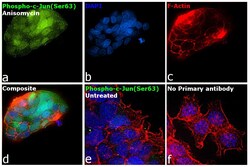
- Immunofluorescence analysis of Transcription factor AP-1 was performed using 70% confluent log phase HEK-293 cells treated with Anisomycin (10 µg/mL for 30 mins). The cells were fixed with 4% paraformaldehyde for 5 minutes, permeabilized with 0.1% Triton™ X-100 for 10 minutes, and blocked with 2% BSA for 45 minutes at room temperature. The cells were labeled with Phospho-c-Jun (Ser63) Polyclonal Antibody (Product # PA5-17890) at 1:100 dilution in 0.1% BSA, incubated at 4 degree celsius overnight and then labeled with Donkey anti-Rabbit IgG (H+L) Highly Cross-Adsorbed Secondary Antibody, Alexa Fluor Plus 488 (Product # A32790), (1:2000 dilution), for 45 minutes at room temperature (Panel a: Green). Nuclei (Panel b:Blue) were stained with ProLong™ Diamond Antifade Mountant with DAPI (Product # P36962). F-actin (Panel c: Red) was stained with Rhodamine Phalloidin (Product # R415, 1:300). Panel d represents the merged image showing nucleus and cytoplasm localization. Panel e represents untreated control HEK-293 cells. Panel f represents control cells with no primary antibody to assess background. The images were captured at 60X magnification.

- Submitted by
- Invitrogen Antibodies (provider)
- Main image

- Experimental details
- Immunofluorescent analysis of Phospho-c-Jun pSer63 II in HeLa cells, anisomycin-treated, using a Phospho-c-Jun pSer63 II polyclonal antibody (Product # PA5-17890) (green). Actin filaments are labeled with a fluorescent red phalloidin. DNA is labeled using a fluorescent blue dye.

- Submitted by
- Invitrogen Antibodies (provider)
- Main image

- Experimental details
- Immunofluorescence analysis of Transcription factor AP-1 was performed using 70% confluent log phase HEK-293 cells treated with Anisomycin (10 µg/mL for 30 mins). The cells were fixed with 4% paraformaldehyde for 5 minutes, permeabilized with 0.1% Triton™ X-100 for 10 minutes, and blocked with 2% BSA for 45 minutes at room temperature. The cells were labeled with Phospho-c-Jun (Ser63) Polyclonal Antibody (Product # PA5-17890) at 1:100 dilution in 0.1% BSA, incubated at 4 degree celsius overnight and then labeled with Donkey anti-Rabbit IgG (H+L) Highly Cross-Adsorbed Secondary Antibody, Alexa Fluor Plus 488 (Product # A32790), (1:2000 dilution), for 45 minutes at room temperature (Panel a: Green). Nuclei (Panel b:Blue) were stained with ProLong™ Diamond Antifade Mountant with DAPI (Product # P36962). F-actin (Panel c: Red) was stained with Rhodamine Phalloidin (Product # R415, 1:300). Panel d represents the merged image showing nucleus and cytoplasm localization. Panel e represents untreated control HEK-293 cells. Panel f represents control cells with no primary antibody to assess background. The images were captured at 60X magnification.

- Submitted by
- Invitrogen Antibodies (provider)
- Main image

- Experimental details
- Immunofluorescent analysis of Phospho-c-Jun pSer63 II in HeLa cells, anisomycin-treated, using a Phospho-c-Jun pSer63 II polyclonal antibody (Product # PA5-17890) (green). Actin filaments are labeled with a fluorescent red phalloidin. DNA is labeled using a fluorescent blue dye.

Supportive validation
- Submitted by
- Invitrogen Antibodies (provider)
- Main image
- Experimental details
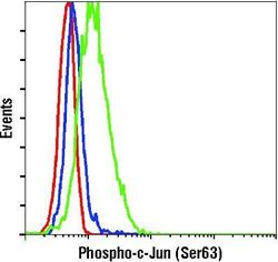
- Flow cytometric analysis of Phospho-c-Jun pSer63 II in untreated (blue) or anisomycin-treated (green) Jurkat cells using a Phospho-c-Jun pSer63 II polyclonal antibody (Product # PA5-17890) compared to a nonspecific negative control antibody (red).

Supportive validation
- Submitted by
- Invitrogen Antibodies (provider)
- Main image
- Experimental details
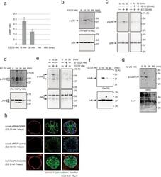
- Figure 2 Analysis of E2 signal transduction. ( a ) cAMP assay showing cAMP levels (nM) in MCF-10A cells following treatment with 32 nM E2 for 15 min, 30 min, 24 h, and 48 h. Three independent experiments were performed. Bars represent +/-SD. ( b ) Western blotting of MCF-10A cells showing p38 and phospho-p38 (Thr180/Tyr182) following treatment with 32 nM E2 for 0-60 min. ( c ) Western blotting of MCF-10A cells treated with 32 nM E2 (left panel) or with 32 nM E2 and 20 nM G-15 (right panel) for 0-30 min. ( d ) Western blotting of MCF-10A cells showing JNK and phosphor-JNK (Thr183/Tyr185) following treatment with 32 nM E2 for 0-60 min. ( e ) Western blotting of MCF-10A cells treated with 32 nM E2 (left panel) or with 32 nM E2 and 20 nM G-15 (right panel) for 0-30 min. ( f ) Western blotting of MCF-10A cells treated with 32 nM E2 for 0-60 min showing IkB and phospho-IkB (Ser32, Ser36). ( g ) Western blotting of MCF-10A cells treated with 32 nM E2 for 0-60 min showing c-Jun and phospho-c-Jun (Ser63). ( h ) Representative confocal images of Accell siRNA-GPER- or siRNA-control-transfected MCF-10A cells in a 3D culture through the middle acini, which were treated with E2 (32 nM, left panels) or control (0 nM, right panel) for 7 days. Laminin V (red); pan-cadherin (green). Scale bars = 20 mum. The presented blots were cropped. Full-length blots are presented in Supplementary Fig. 5 .
