 Explore
 Validate
 Learn
Learn Western blot
 Immunohistochemistry
Antibody data
- Antibody Data
- Antigen structure
- References [0]
- Comments [0]
- Validations
- Immunohistochemistry [2]
Submit
Validation data
Reference
Comment
Report error
- Product number
- PA1-16612 - Provider product page

- Provider
- Invitrogen Antibodies
- Product name
- VEGF Receptor 1/2 Polyclonal Antibody
- Antibody type
- Polyclonal
- Antigen
- Synthetic peptide
- Description
- The target sequence has 100% sequence homology with mouse and rat.
- Reactivity
- Human, Rat
- Host
- Rabbit
- Isotype
- IgG
- Vial size
- 100 μL
- Concentration
- 1 mg/mL
- Storage
- 4°C, do not freeze
No comments: Submit comment
Supportive validation
- Submitted by
- Invitrogen Antibodies (provider)
- Main image
- Experimental details
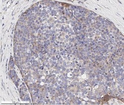
- Immunohistochemical analysis of VEGF Receptor 1/2 in formalin-fixed paraffin-embedded human breast carcinoma tissue. Samples were incubated in VEGF Receptor 1/2 polyclonal antibody (Product # PA1-16612) using a dilution of 1:500. Bond Rx autostainer (Leica Biosystems). The assay involved 20 minutes of heat induced antigen retrieval (HIER) with 10mM sodium citrate buffer (pH 6.0) and endogenous peroxidase quenching using peroxide block. The sections were incubated with primary antibody for 30 minutes. Bond Polymer Refine Detection (Leica Biosystems) and DAB were used for signal detection which followed counterstaining with hematoxylin. Whole slide scanning and capturing of representative images (20X) were performed using Aperio AT2 (Leica Biosystems). This VEGFR1/Flt-1 antibody generated an expected membrane cytoplasmic staining of VEGFR1 protein in the cancer cells (punctate appearance typical of receptors). The tumor stroma/stromal cells did not show VEGFR1/Flt-1immunopositivity.

- Submitted by
- Invitrogen Antibodies (provider)
- Main image
- Experimental details
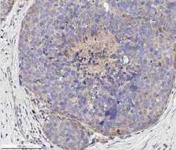
- Immunohistochemical analysis of VEGF Receptor 1/2 in formalin-fixed paraffin-embedded human breast carcinoma tissue section. Samples were incubated in VEGF Receptor 1/2 polyclonal antibody (Product # PA1-16612) using a dilution of 1:500. Bond Rx autostainer (Leica Biosystems). The assay involved 20 minutes of heat induced antigen retrieval (HIER) with 10mM sodium citrate buffer (pH 6.0) and endogenous peroxidase quenching using peroxide block. The sections were incubated with primary antibody for 30 minutes. Bond Polymer Refine Detection (Leica Biosystems) and DAB were used for signal detection which followed counterstaining with hematoxylin. Whole slide scanning and capturing of representative images (20X) were performed using Aperio AT2 (Leica Biosystems). This VEGFR1/Flt-1 antibody generated an expected membrane cytoplasmic staining of VEGFR1 protein in the cancer cells. The tumor stroma/stromal cells did not show VEGFR1/Flt-1immunopositivity. Staining was performed by Histowiz.
