 Explore
 Validate
 Learn
LearnMA1-16757
antibody from Invitrogen Antibodies
Targeting: GAPDHS
GAPD2, GAPDH-2, GAPDS
 Western blot Immunocytochemistry
 Immunoprecipitation Immunohistochemistry Chromatin Immunoprecipitation Other assay
Antibody data
- Antibody Data
- Antigen structure
- References [0]
- Comments [0]
- Validations
- Western blot [5]
- Immunocytochemistry [4]
- Other assay [9]
Submit
Validation data
Reference
Comment
Report error
- Product number
- MA1-16757 - Provider product page

- Provider
- Invitrogen Antibodies
- Product name
- GAPDH Monoclonal Antibody (1D4)
- Antibody type
- Monoclonal
- Antigen
- Purifed from natural sources
- Description
- This antibody is likely to react with most mammals. Suggested positive control: antigen standard for GAPDH (transient overexpression lysate).
- Reactivity
- Human, Mouse, Rat, Bovine, Canine, Chicken/Avian, Drosophila, Feline, Hamster, Porcine, Rabbit, Zebrafish
- Host
- Mouse
- Isotype
- IgM
- Antibody clone number
- 1D4
- Vial size
- 100 μL
- Concentration
- 1 mg/mL
- Storage
- Store at 4°C short term. For long term storage, store at -20°C, avoiding freeze/thaw cycles.
No comments: Submit comment
Supportive validation
- Submitted by
- Invitrogen Antibodies (provider)
- Main image
- Experimental details
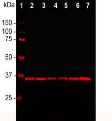
- Western blot analysis of GAPDH in cell line lysates. Samples were incubated in GAPDH monoclonal antibody (Product # MA1-16757 using a dilution of 1:2,000. [1] protein standard, [2] HEK293, [3] HeLa, [4] SH-SY5Y, [5] COS1, [6] NIH-3T3, and [7] C6 cells. The GAPDH antibody reveals a single band at ~37 kDa in all cell lines. GAPDH is a house keeping protein, the level of which is relatively unaffected by most experimental manipulations, and, as a result, this antibody has been widely used as a ting loading control.

- Submitted by
- Invitrogen Antibodies (provider)
- Main image
- Experimental details
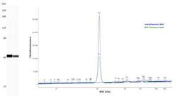
- Western blot analysis of GAPDH in 0.05 mg/mL Jurkat and MCF-7 cell lysates. Samples were incubated in GAPDH monoclonal antibody (Product # MA1-16757) using a dilution of 1:25. Electropherogram image of corresponding Simple Western lanes view at WES molecular weight of 40 kDa.

- Submitted by
- Invitrogen Antibodies (provider)
- Main image
- Experimental details
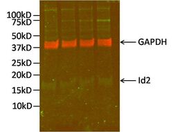
- Western blot analysis of GAPDH was performed by loading 10 µg of Id2-transfected SK-N-BE human neuroblastoma cells (in replicate) per well onto an SDS-PAGE gel. Proteins were transferred to a PVDF membrane and blocked for 1 hour. The membrane was probed with a GAPDH monoclonal antibody (Product # MA5-16757) at a dilution of 1:2000 overnight at 4ºC, washed in 0.1% PBS-Tween, and probed with an infrared dye-conjugated anti-mouse IgG secondary antibody (shown in red) at a dilution of 1:20,000 for 1 hour. The blot was also probed with an Id2 monoclonal antibody (Product # MA5-14777) at a dilution of 1:500 followed by an infrared Dye-conjugated anti-rabbit IgG secondary antibody (shown in green) at a dilution of 1:15,000. Detection was performed using a fluorescent scanner. Data courtesy of the Innovators Program.

- Submitted by
- Invitrogen Antibodies (provider)
- Main image
- Experimental details
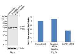
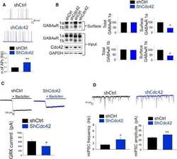
- Knockdown of GAPDH was achieved by transfecting HeLa cells with GAPDH specific validated siRNAs (Silencer® select Product # s13960 ). Western blot analysis (Fig. a) was performed using whole cell extracts from the GAPDH knockdown cells (lane 3), non-specific scrambled siRNA transfected cells (lane 2) and untransfected cells (lane 1). The blots were probed with GAPDH Monoclonal Antibody (Product # MA1-16757, 1:2000 dilution) and Goat anti-Mouse IgG (H+L) Superclonal™ Secondary Antibody, HRP conjugate (Product # A28177, 0.25 µg/mL, 1:4000 dilution). Densitometric analysis of this western blot is shown in histogram (Fig. b). Decrease in signal upon siRNA mediated knock down confirms that antibody is specific to GAPDH.

- Submitted by
- Invitrogen Antibodies (provider)
- Main image
- Experimental details
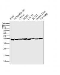
- Western blot analysis was performed on whole cell extracts (30 µg lysate) of A549 (Lane 1), MDA-MB-231 (Lane 2), COS-7 (Lane 3), MDCK (Lane 4), C2C12 (Lane 5), PC-12 (Lane 6) and tissue extracts (30 µg lysate) of Mouse Liver (Lane 7) and Rat Lung (Lane 8 ). The blot was probed with Anti-GAPDH Monoclonal Antibody (Product # MA1-16757, 1:1000 dilution) and detected by chemiluminescence using Goat anti-Mouse IgG (H+L) Superclonal™ Secondary Antibody, HRP conjugate (Product # A28177, 0.25 µg/mL, 1:4000 dilution). A 37 kDa band corresponding to GAPDH was observed across the cell lines and tissue extracts tested.

Supportive validation
- Submitted by
- Invitrogen Antibodies (provider)
- Main image

- Experimental details
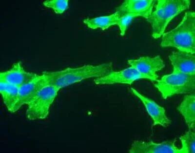
- Immunocytochemistry analysis of GAPDH in HeLa cells. Samples were incubated in GAPDH monoclonal antibody (Product # MA1-16757) using a dilution of 1:100. Antibody (Green). DAPI staining of nuclear DNA (Blue). The GAPDHantibody produces strong cytoplasmic staining of healthy cells.

- Submitted by
- Invitrogen Antibodies (provider)
- Main image

- Experimental details
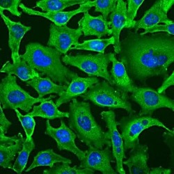
- Immunocytochemistry analysis of GAPDH in SH-SY5Y cells. Samples were incubated in GAPDH monoclonal antibody (Product # MA1-16757). GAPDH antibody (green). Nuclear DNA is stained with Hoechst dye (blue).
- Submitted by
- Invitrogen Antibodies (provider)
- Main image
- Experimental details
- Immunocytochemistry analysis of GAPDH in HeLa cells. Samples were incubated in GAPDH monoclonal antibody (Product # MA1-16757) using a dilution of 1:100. Antibody (Green). DAPI staining of nuclear DNA (Blue). The GAPDHantibody produces strong cytoplasmic staining of healthy cells.

- Submitted by
- Invitrogen Antibodies (provider)
- Main image

- Experimental details
- Immunocytochemistry analysis of GAPDH in SH-SY5Y cells. Samples were incubated in GAPDH monoclonal antibody (Product # MA1-16757). GAPDH antibody (green). Nuclear DNA is stained with Hoechst dye (blue).

Supportive validation
- Submitted by
- Invitrogen Antibodies (provider)
- Main image
- Experimental details
- NULL

- Submitted by
- Invitrogen Antibodies (provider)
- Main image
- Experimental details
- NULL

- Submitted by
- Invitrogen Antibodies (provider)
- Main image

- Experimental details
- NULL
- Submitted by
- Invitrogen Antibodies (provider)
- Main image
- Experimental details
- NULL
- Submitted by
- Invitrogen Antibodies (provider)
- Main image
- Experimental details
- NULL
- Submitted by
- Invitrogen Antibodies (provider)
- Main image
- Experimental details
- NULL
- Submitted by
- Invitrogen Antibodies (provider)
- Main image
- Experimental details
- NULL

- Submitted by
- Invitrogen Antibodies (provider)
- Main image
- Experimental details
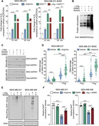
- Fig. 5. l -Arginine generates peroxynitrate, GAPDH nitrosylation, and protein ADP-ribosylation. ( A ) Peroxynitrate levels in MDA-MB-231 and MDA-MB-231-BrM2 cells treated with vehicle, l -arginine, the SOD mimetic tempol (SOD1 mim ), or the combination of l -arginine and SOD1 mim for 15 and 55 min. ( B ) Total protein nitrosylation in MDA-MB-231 cells exposed to l -arginine for 60 min. GSNO was used as a positive control. ( C ) SNO-GAPDH (and total GAPDH) in MDA-MB-231 and MDA-MB-468 cells exposed for 60 min to l -arginine alone or in combination with the NOS2 inhibitor 1400W. GSNO was used as a positive control. ( D ) DNA damage levels assessed by comet assay in MDA-MB-231 and MDA-MB-231-BrM2 cells exposed to vehicle or l -arginine for 60 min with and without Fpg. ( E ) Total mono- and poly-ADP-ribosylated proteins in MDA-MB-231 and MDA-MB-468 cells exposed to l -arginine alone or in combination with the NOS2 inhibitor 1400W. ( F ) NAD + levels in MDA-MB-231 and MDA-MB-468 cells exposed to l -arginine alone or in combination with the PARP inhibitor olaparib. ** P < 0.001 and *** P < 0.0001.

- Submitted by
- Invitrogen Antibodies (provider)
- Main image
- Experimental details
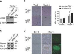
- Figure 1 The effect of ASB6 overexpression on soft agar colony formation, stemness genes expression, and tumor sphere formation of OSCC cells. The OECM1 cells with stable overexpression of green fluorescent protein (vector GFP) or GFP-tagged ASB6 (ASB6-GFP) were validated by western blot for ASB6 (with as beta-actin as the loading control) (A) , and were subjected to anchorage-independent growth analysis by the soft agar assay (B) , western blots analysis for Nanog and Oct-4 (with as the GAPDH as loading control) (C) , and tumor sphere formation analysis (D) .* P < 0.05.
