Explore
 Validate
 Learn
Learn Western blot
 Immunocytochemistry
Antibody data
- Antibody Data
- Antigen structure
- References [1]
- Comments [0]
- Validations
- Immunocytochemistry [1]
- Immunohistochemistry [2]
- Other assay [1]
Submit
Validation data
Reference
Comment
Report error
- Product number
- PA5-20721 - Provider product page

- Provider
- Invitrogen Antibodies
- Product name
- ApoA1 Polyclonal Antibody
- Antibody type
- Polyclonal
- Antigen
- Synthetic peptide
- Description
- A suggested positive control is human liver tissue lysate. PA5-20721 can be used with blocking peptide PEP-0835.
- Reactivity
- Human, Mouse, Rat
- Host
- Chicken/Avian
- Isotype
- IgY
- Vial size
- 100 μg
- Concentration
- 1 mg/mL
- Storage
- Maintain refrigerated at 2-8°C for up to 3 months. For long term storage store at -20°C
Submitted references Activation of neutral sphingomyelinase 2 through hyperglycemia contributes to endothelial apoptosis via vesicle-bound intercellular transfer of ceramides.
Zietzer A, Jahnel AL, Bulic M, Gutbrod K, Düsing P, Hosen MR, Dörmann P, Werner N, Nickenig G, Jansen F
Cellular and molecular life sciences : CMLS 2021 Dec 24;79(1):48
Cellular and molecular life sciences : CMLS 2021 Dec 24;79(1):48
No comments: Submit comment
Supportive validation
- Submitted by
- Invitrogen Antibodies (provider)
- Main image
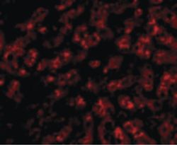
- Experimental details
- Immunofluorescent analysis of human liver cells using a ApoA1 polyclonal antibody (Product # PA5-20721) at a 20 µg/mL dilution.

Supportive validation
- Submitted by
- Invitrogen Antibodies (provider)
- Main image
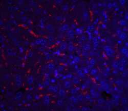
- Experimental details
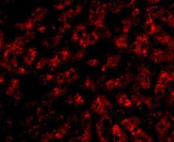
- Immunofluorescence of ApoA1 in mouse liver tissue with ApoA1 Polyclonal Antibody (Product # PA5-20721) at 20 µg/mL. Red: ApoA1 Blue: DAPI staining
- Submitted by
- Invitrogen Antibodies (provider)
- Main image
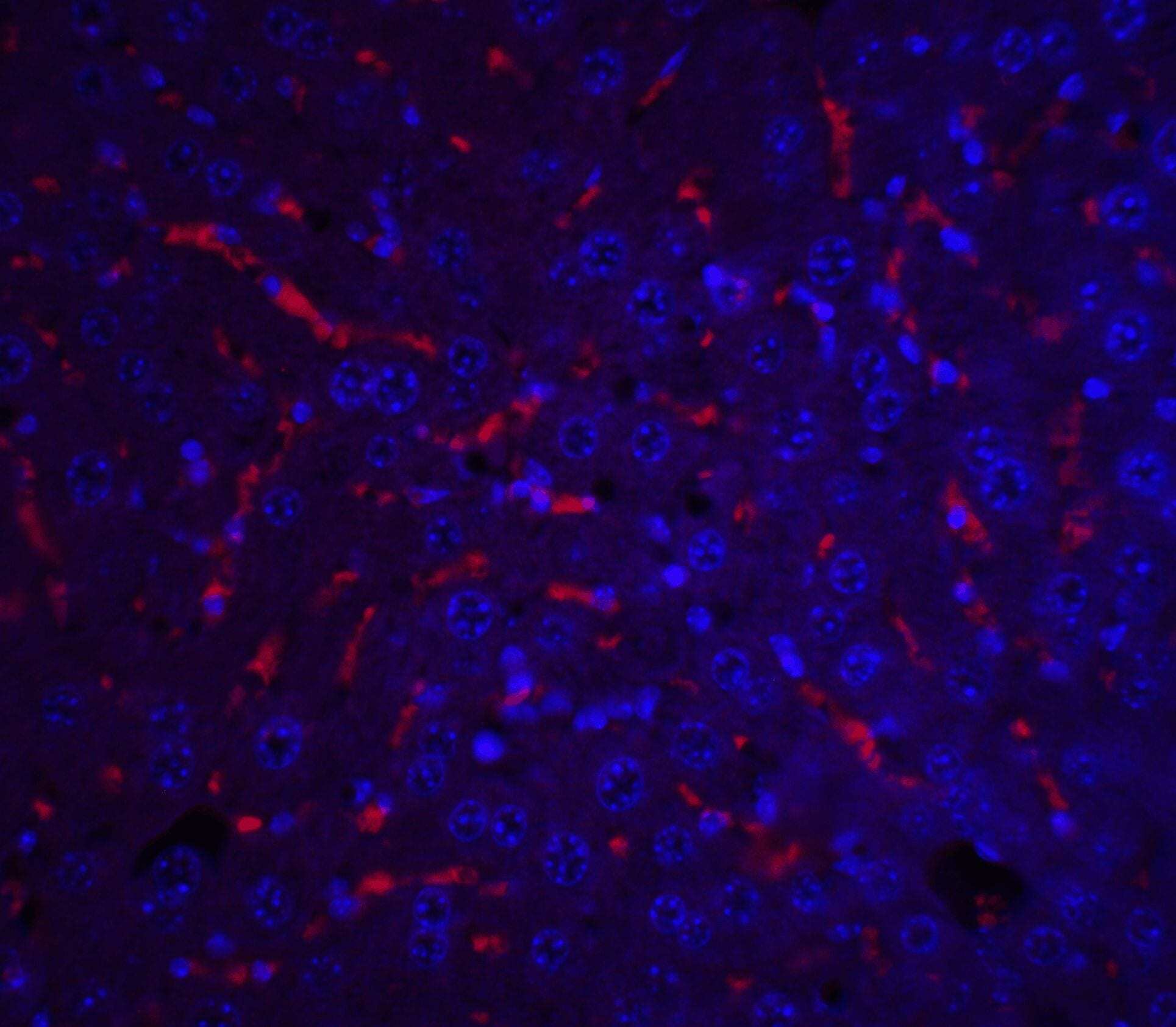
- Experimental details
- Immunofluorescence of ApoA1 in human liver tissue with ApoA1 Polyclonal Antibody (Product # PA5-20721) at 20 µg/mL.

Supportive validation
- Submitted by
- Invitrogen Antibodies (provider)
- Main image
- Experimental details
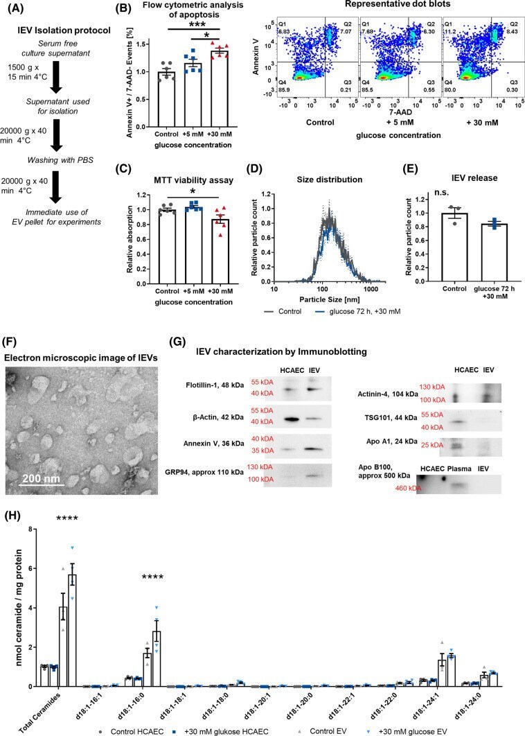
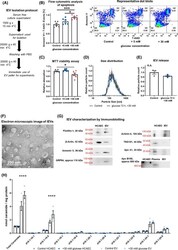
- Fig. 1 A Schematic diagram of the lEV isolation protocol. B Flow-cytometric analysis of apoptosis induction in HCAECs after hyperglycemic injury, n = 6, with representative dot blots. C MTT viability assay in HCAECs after hyperglycemic injury, n = 6. D + E Distribution of lEV size and relative lEVs released after hyperglycemic injury in HCAECs, as analyzed by nanoparticle tracking analysis, n = 3. F Direct electron microscopic imaging of HCAEC-derived lEVs with negative staining, representative image. G Characterization of lEVs by immunoblotting for Flotillin-1, beta -Actin, Annexin V, GRP94, Actinin-4, TSG101, Apolipoprotein A1, Apolipoprotein B100 (with additional human plasma as an antibody control). H Mass spectrometric analysis of ceramides in HCAECs and lEVs after hyperglycemic injury. All data are presented as individual experiments with the mean +- SEM; n.s. not significant, * p < 0.05, *** p < 0.001, **** p < 0.0001. ANOVA + Bonferoni's multiple comparison test were used for B + C , unpaired t -Test for E , two-way ANOVA + Bonferoni's multiple comparison test were used for H