Explore
 Validate
 Learn
Learn Western blot
 Immunocytochemistry
Antibody data
- Antibody Data
- Antigen structure
- References [0]
- Comments [0]
- Validations
- Immunocytochemistry [1]
- Immunohistochemistry [2]
Submit
Validation data
Reference
Comment
Report error
- Product number
- PA5-32163 - Provider product page

- Provider
- Invitrogen Antibodies
- Product name
- SERPINE2 Polyclonal Antibody
- Antibody type
- Polyclonal
- Antigen
- Recombinant full-length protein
- Description
- Recommended positive controls: HeLa, HepG2, mouse brain. Predicted reactivity: Mouse (87%), Rat (87%), Pig (94%), Chicken (82%), Bovine (93%). PA5-32163 detects SERPINE2 protein in formalin-fixed paraffin-embedded equine endometrial tissue. Store product as a concentrated solution. Centrifuge briefly prior to opening the vial.
- Reactivity
- Human, Mouse
- Host
- Rabbit
- Isotype
- IgG
- Vial size
- 100 μL
- Concentration
- 1 mg/mL
- Storage
- Store at 4°C short term. For long term storage, store at -20°C, avoiding freeze/thaw cycles.
No comments: Submit comment
Supportive validation
- Submitted by
- Invitrogen Antibodies (provider)
- Main image
- Experimental details
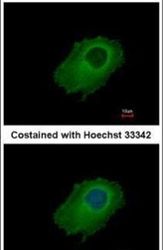
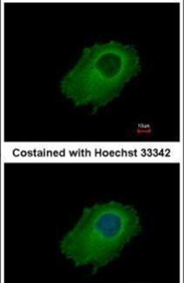
- Immunofluorescent analysis of SERPINE2 in methanol-fixed HeLa cells using a SERPINE2 polyclonal antibody (Product # PA5-32163) at a 1:500 dilution.
Supportive validation
- Submitted by
- Invitrogen Antibodies (provider)
- Main image
- Experimental details
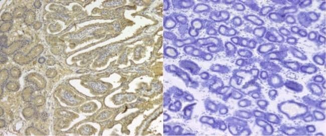
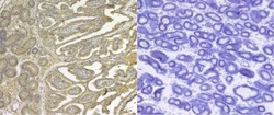
- Immunohistochemical analysis of paraffin-embedded human gastric cancer, using SERPINE2 (Product # PA5-32163) antibody at 1:500 dilution. Antigen Retrieval: EDTA based buffer, pH 8.0, 15 min.
- Submitted by
- Invitrogen Antibodies (provider)
- Main image
- Experimental details
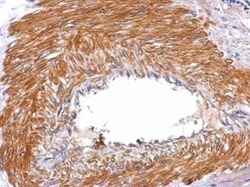
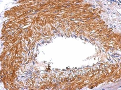
- Immunohistochemistry analysis of SERPINE2 was performed on equine endometrial tissue sections. 5-µm-thick sections were prepared from formalin-fixed paraffin-embedded tissue blocks and were dried on slide plate at 37°C overnight. The sections were placed in a Bond Max Automated Immunohistochemistry Vision Biosystem (Leica Microsystems GmbH, Wetzlar, Germany) according to the following protocol. First, tissues were deparaffinized and pre-treated with Epitope Retrieval Solution 2 (Citrate based pH8) at 98°C for 10 minutes. After washing steps, peroxidase blocking was carried out for 10 min using the Bond Polymer Refine Detection Kit DC9800 (Leica Microsystems GmbH). Tissues were again washed and then incubated with the primary antibody (Product # PA5-32163, 1:200) for 30 min then the secondary antibody diluted 1:1000. Subsequently, tissues were incubated with polymer for 10 min and developed with DAB-Chromogen for 10 min. Data courtesy of Antibody Data Exchange Program.