Explore
 Validate
 Learn
LearnMAB226-100
antibody from R&D Systems
Targeting: TNFRSF1B
CD120b, p75, TNF-R-II, TNF-R75, TNFBR, TNFR2, TNFR80
 Immunocytochemistry
Antibody data
- Antibody Data
- Antigen structure
- References [6]
- Comments [0]
- Validations
- Immunocytochemistry [1]
- Flow cytometry [1]
- Blocking/Neutralizing [1]
Submit
Validation data
Reference
Comment
Report error
- Product number
- MAB226-100 - Provider product page

- Provider
- R&D Systems
- Product name
- Human TNF RII/TNFRSF1B Antibody
- Antibody type
- Monoclonal
- Description
- Protein A or G purified from ascites. Detects human TNF RII/TNFRSF1B in direct ELISAs. In direct ELISAs, no cross-reactivity with recombinant human (rh) 4-1BB, rhCD27, rhCD30, rhCD40, rhDR3, rhDR6, rhFas, rhGITR, rhHVEM, rhLTR beta , rhNGF R, rhOPG, rhRANK, rhTNF RI, or recombinant mouse TNF RII is observed. Neutralizes the biological effect of rhTNF RII but not rhTNF RI.
- Reactivity
- Human
- Host
- Mouse
- Conjugate
- Unconjugated
- Antigen sequence
P20333- Isotype
- IgG
- Antibody clone number
- 22221
- Vial size
- 100 ug
- Storage
- Use a manual defrost freezer and avoid repeated freeze-thaw cycles. 12 months from date of receipt, -20 to -70 °C as supplied. 1 month, 2 to 8 °C under sterile conditions after reconstitution. 6 months, -20 to -70 °C under sterile conditions after reconstitution.
Submitted references Low doses of LPS exacerbate the inflammatory response and trigger death on TLR3-primed human monocytes.
Tumor necrosis factor stimulates osteoclastogenesis from human bone marrow cells under hypoxic conditions.
FOXO3a regulates oxygen-responsive expression of tumor necrosis factor receptor 2 in human dermal microvascular endothelial cells.
Cutting edge: TNFR-shedding by CD4+CD25+ regulatory T cells inhibits the induction of inflammatory mediators.
Human intestinal intraepithelial lymphocytes keep TNF alpha levels low by cell uptake and feedback inhibition of transcription.
Synergistic induction of apoptosis in breast cancer cells by cotreatment with butyrate and TNF-alpha, TRAIL, or anti-Fas agonist antibody involves enhancement of death receptors' signaling and requires P21(waf1).
Monguió-Tortajada M, Franquesa M, Sarrias MR, Borràs FE
Cell death & disease 2018 May 1;9(5):499
Cell death & disease 2018 May 1;9(5):499
Tumor necrosis factor stimulates osteoclastogenesis from human bone marrow cells under hypoxic conditions.
Nomura T, Aoyama M, Waguri-Nagaya Y, Goto Y, Suzuki M, Miyazawa K, Asai K, Goto S
Experimental cell research 2014 Feb 15;321(2):167-77
Experimental cell research 2014 Feb 15;321(2):167-77
FOXO3a regulates oxygen-responsive expression of tumor necrosis factor receptor 2 in human dermal microvascular endothelial cells.
Ding B, Kirkiles-Smith NC, Pober JS
The Journal of biological chemistry 2009 Jul 17;284(29):19331-9
The Journal of biological chemistry 2009 Jul 17;284(29):19331-9
Cutting edge: TNFR-shedding by CD4+CD25+ regulatory T cells inhibits the induction of inflammatory mediators.
van Mierlo GJ, Scherer HU, Hameetman M, Morgan ME, Flierman R, Huizinga TW, Toes RE
Journal of immunology (Baltimore, Md. : 1950) 2008 Mar 1;180(5):2747-51
Journal of immunology (Baltimore, Md. : 1950) 2008 Mar 1;180(5):2747-51
Human intestinal intraepithelial lymphocytes keep TNF alpha levels low by cell uptake and feedback inhibition of transcription.
Ebert EC, Mehta V
Cellular immunology 2006 May;241(1):7-13
Cellular immunology 2006 May;241(1):7-13
Synergistic induction of apoptosis in breast cancer cells by cotreatment with butyrate and TNF-alpha, TRAIL, or anti-Fas agonist antibody involves enhancement of death receptors' signaling and requires P21(waf1).
Chopin V, Slomianny C, Hondermarck H, Le Bourhis X
Experimental cell research 2004 Aug 15;298(2):560-73
Experimental cell research 2004 Aug 15;298(2):560-73
No comments: Submit comment
Supportive validation
- Submitted by
- R&D Systems (provider)
- Main image

- Experimental details
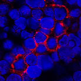
- TNF RII/TNFRSF1B in Human PBMCs. TNF RII/TNFRSF1B was detected in immersion fixed human peripheral blood mononuclear cells (PBMCs) using Mouse Anti-Human TNF RII/TNFRSF1B Monoclonal Antibody (Catalog # MAB226) at 25 µg/mL for 3 hours at room temperature. Cells were stained using the NorthernLights™ 557-conjugated Anti-Mouse IgG Secondary Antibody (red; Catalog # NL007) and counterstained with DAPI (blue). Specific staining was localized to plasma membrane and cytoplasm. View our protocol for Fluorescent ICC Staining of Cells on Coverslips.
Supportive validation
- Submitted by
- R&D Systems (provider)
- Main image

- Experimental details
- Detection of TNF RII/TNFRSF1B in Human Granulocytes by Flow Cytometry. Human peripheral blood granulocytes were stained with Mouse Anti-Human TNF RII/TNFRSF1B Monoclonal Antibody (Catalog # MAB226, filled histogram) or isotype control antibody (Catalog # MAB003, open histogram) followed by anti-mouse IgG PE-conjugated secondary antibody (Catalog # F0101B). View our protocol for Staining Membrane-associated Proteins.

Supportive validation
- Submitted by
- R&D Systems (provider)
- Main image

- Experimental details
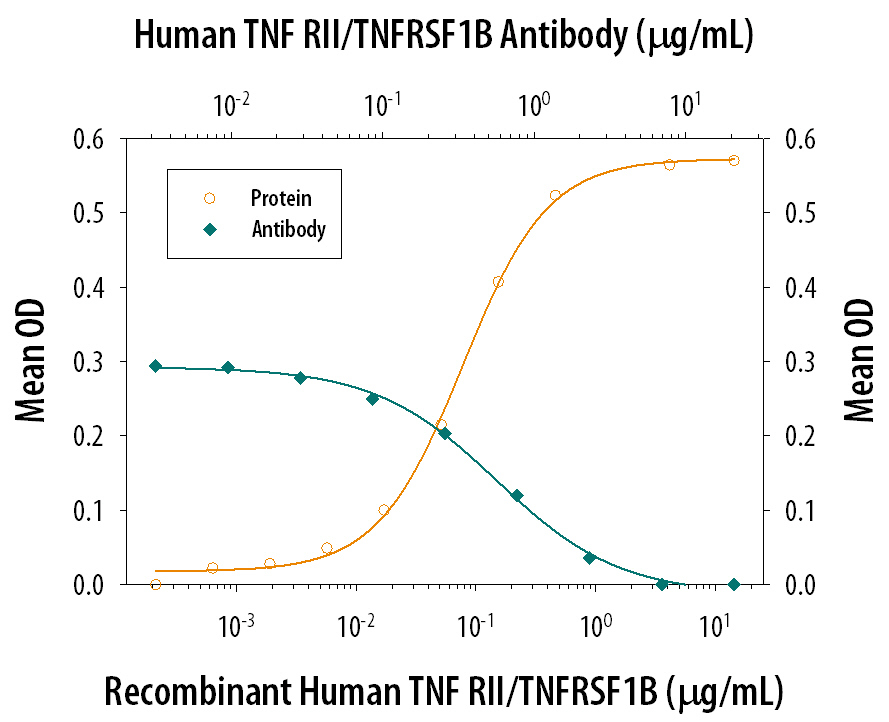
- TNF RII/TNFRSF1B Inhibition of TNF-alpha-induced Cytotoxicity and Neutralization by Human TNF RII/TNFRSF1B Antibody. Recombinant Human TNF RII/TNFRSF1B inhibits Recombinant Human TNF-alpha (Catalog # 210-TA) induced cytotoxicity in the L-929 mouse fibroblast cell line in a dose-dependent manner (orange line), as measured by crystal violet staining. Inhibition of Recombinant Human TNF-alpha (0.25 ng/mL) activity elicited by Recombinant Human TNF RII/ TNFRSF1B (0.3 µg/mL) is neutralized (green line) by increasing concentrations of Mouse Anti-Human TNF RII/ TNFRSF1B Monoclonal Antibody (Catalog # MAB226). The ND50 is typically 0.5-1.5 µg/mL in the presence of the metabolic inhibitor actinomycin D (1 µg/mL).