Explore
 Validate
 Learn
Learn Western blot
 Immunocytochemistry
Antibody data
- Antibody Data
- Antigen structure
- References [0]
- Comments [0]
- Validations
- Immunocytochemistry [1]
- Immunohistochemistry [1]
- Flow cytometry [1]
Submit
Validation data
Reference
Comment
Report error
- Product number
- AF3587 - Provider product page

- Provider
- R&D Systems
- Product name
- Human Carboxypeptidase E/CPE Antibody
- Antibody type
- Polyclonal
- Description
- Antigen Affinity-purified. Detects human Carboxypeptidase E/CPE in direct ELISAs and Western blots. In direct ELISAs, less than 5% cross-reactivity with recombinant human CPM is observed.
- Reactivity
- Human
- Host
- Goat
- Conjugate
- Unconjugated
- Antigen sequence
P16870- Isotype
- IgG
- Vial size
- 100 ug
- Concentration
- LYOPH
- Storage
- Use a manual defrost freezer and avoid repeated freeze-thaw cycles. 12 months from date of receipt, -20 to -70 °C as supplied. 1 month, 2 to 8 °C under sterile conditions after reconstitution. 6 months, -20 to -70 °C under sterile conditions after reconstitution.
No comments: Submit comment
Supportive validation
- Submitted by
- R&D Systems (provider)
- Main image
- Experimental details
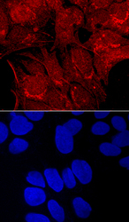
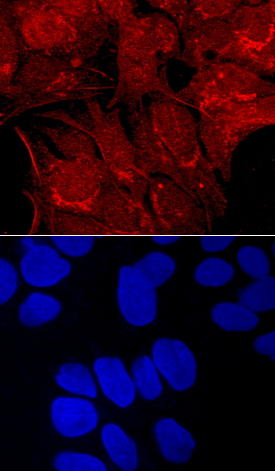
- Carboxypeptidase E/CPE in HepG2 Human Cell Line. Carboxypeptidase E/CPE was detected in immersion fixed HepG2 human hepatocellular carcinoma cell line using Goat Anti-Human Carboxypeptidase E/CPE Antigen Affinity-purified Polyclonal Antibody (Catalog # AF3587) at 10 µg/mL for 3 hours at room temperature. Cells were stained using the NorthernLights™ 557-conjugated Anti-Goat IgG Secondary Antibody (red, upper panel; Catalog # NL001) and counterstained with DAPI (blue, lower panel). Specific staining was localized to cell surfaces and cytoplasm. View our protocol for Fluorescent ICC Staining of Cells on Coverslips.
Supportive validation
- Submitted by
- R&D Systems (provider)
- Main image
- Experimental details
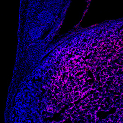
- Carboxypeptidase E/CPE in Mouse Embryo. Carboxypeptidase E/CPE was detected in immersion fixed frozen sections of mouse embryo (9.5 d.p.c.) using Goat Anti-Human Carboxypeptidase E/CPE Antigen Affinity-purified Polyclonal Antibody (Catalog # AF3587) at 10 µg/mL overnight at 4 °C. Tissue was stained using the NorthernLights™ 557-conjugated Anti-Goat IgG Secondary Antibody (red; Catalog # NL001) and counterstained with DAPI (blue). Specific staining was localized to the developing liver. View our protocol for Fluorescent IHC Staining of Frozen Tissue Sections.

Supportive validation
- Submitted by
- R&D Systems (provider)
- Main image

- Experimental details
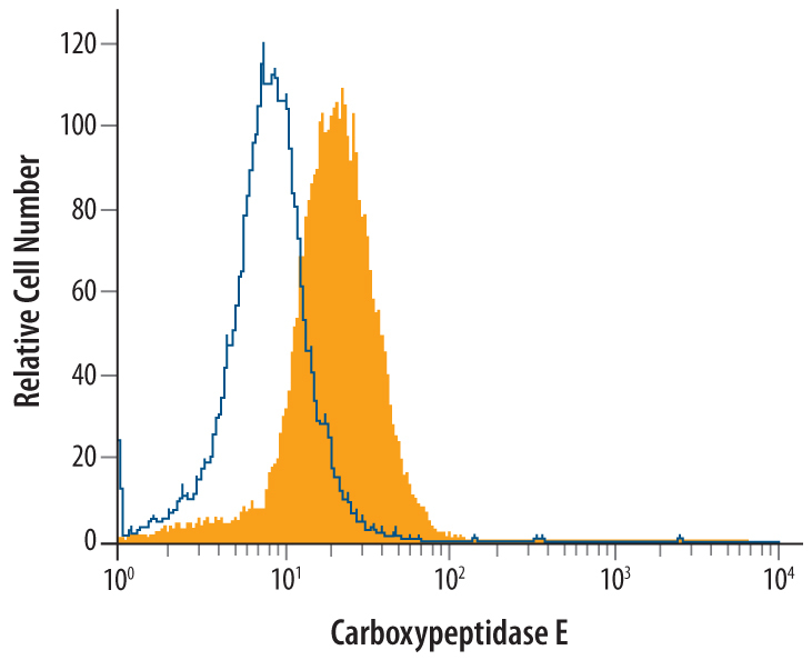
- Detection of Carboxypeptidase E/CPE in A172 Human Cell Line by Flow Cytometry. A172 human glioblastoma cell line was stained with Goat Anti-Human Carboxypeptidase E/CPE Antigen Affinity-purified Polyclonal Antibody (Catalog # AF3587, filled histogram) or control antibody (Catalog # AB-108-C, open histogram), followed by Phycoerythrin-conjugated Anti-Goat IgG Secondary Antibody (Catalog # F0107). To facilitate intracellular staining, cells were fixed with paraformaldehyde and permeabilized with methanol and saponin.