Explore
 Validate
 Learn
Learn Western blot
Antibody data
- Antibody Data
- Antigen structure
- References [1]
- Comments [0]
- Validations
- Western blot [3]
- Immunocytochemistry [4]
- Other assay [1]
Submit
Validation data
Reference
Comment
Report error
- Product number
- MA5-15389 - Provider product page

- Provider
- Invitrogen Antibodies
- Product name
- Calnexin Monoclonal Antibody (3H4A7)
- Antibody type
- Monoclonal
- Antigen
- Synthetic peptide
- Description
- MA5-15389 targets Calnexin in IF and WB applications and shows reactivity with Human samples.
- Antibody clone number
- 3H4A7
- Concentration
- Conc. Not Determined
Submitted references Plasma extracellular vesicles in people living with HIV and type 2 diabetes are related to microbial translocation and cardiovascular risk.
Vestad B, Nyman TA, Hove-Skovsgaard M, Stensland M, Hoel H, Trøseid AS, Aspelin T, Aass HCD, Puhka M, Hov JR, Nielsen SD, Øvstebø R, Trøseid M
Scientific reports 2021 Nov 9;11(1):21936
Scientific reports 2021 Nov 9;11(1):21936
No comments: Submit comment
Supportive validation
- Submitted by
- Invitrogen Antibodies (provider)
- Main image
- Experimental details
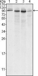
- Western blot analysis of Calnexin using Calnexin monoclonal antibody (Product # MA5-15389) in A431 (1), HeLa (2), MCF-7 (3) and A549 (4) cell lysate.

- Submitted by
- Invitrogen Antibodies (provider)
- Main image

- Experimental details
- Western blot was performed using Anti-Calnexin Monoclonal Antibody (3H4A7) (Product # MA5-15389) and a ~68-75 kDa band corresponding to CANX was observed across cell lines tested . Tissue extracts (30 µg lysate) of HeLa (Lane 1), HCT 116 (Lane 2), A-431 (Lane 3), Hep G2 (Lane 4), Jurkat (Lane 5), RAW 264.7 (Lane 6) were electrophoresed using NuPAGE™ 4-12% Bis-Tris Protein Gel (Product # NP0321BOX). Resolved proteins were then transferred onto a nitrocellulose membrane (Product # IB23001) by iBlot® 2 Dry Blotting System (Product # IB21001). The blot was probed with the primary antibody (1:1000) and detected by chemiluminescence with Goat anti-Mouse IgG (H+L) Superclonal™ Recombinant Secondary Antibody, HRP (Product # A28177,1:20000) using the iBright™ FL1500 Imaging System (Product # A44115). Chemiluminescent detection was performed using SuperSignal™ West Atto Ultimate Sensitivity Substrate (Product # A38556).

- Submitted by
- Invitrogen Antibodies (provider)
- Main image
- Experimental details
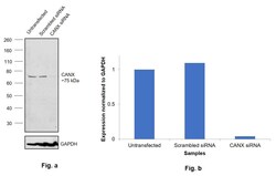
- Knockdown of CANX was achieved by transfecting HeLa with CANX specific siRNAs (Silencer® select Product # s2376, s2378). Western blot analysis (Fig. a) was performed using Whole cell extracts from the CANX knockdown cells (lane 3), non-targeting scrambled siRNA transfected cells (lane 2) and untransfected cells (lane 1). The blot was probed with Calnexin Monoclonal Antibody (3H4A7) (Product # MA5-15389, 1:1000 ) and Goat anti-Mouse IgG (H+L) Superclonal™ Recombinant Secondary Antibody, HRP (Product # A28177, 1:20000). Densitometric analysis of this western blot is shown in histogram (Fig. b). Decrease in signal upon siRNA mediated knock down confirms that antibody is specific to CANX.

Supportive validation
- Submitted by
- Invitrogen Antibodies (provider)
- Main image

- Experimental details
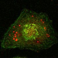
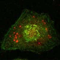
- immunofluorescence analysis of HeLa cells using Calnexin monoclonal antibody (Product # MA5-15389) (green).
- Submitted by
- Invitrogen Antibodies (provider)
- Main image

- Experimental details
- immunofluorescence analysis of HeLa cells using Calnexin monoclonal antibody (Product # MA5-15389) (green).

- Submitted by
- Invitrogen Antibodies (provider)
- Main image

- Experimental details
- immunofluorescence analysis of HeLa cells using Calnexin monoclonal antibody (Product # MA5-15389) (green).
- Submitted by
- Invitrogen Antibodies (provider)
- Main image

- Experimental details
- immunofluorescence analysis of HeLa cells using Calnexin monoclonal antibody (Product # MA5-15389) (green).

Supportive validation
- Submitted by
- Invitrogen Antibodies (provider)
- Main image
- Experimental details
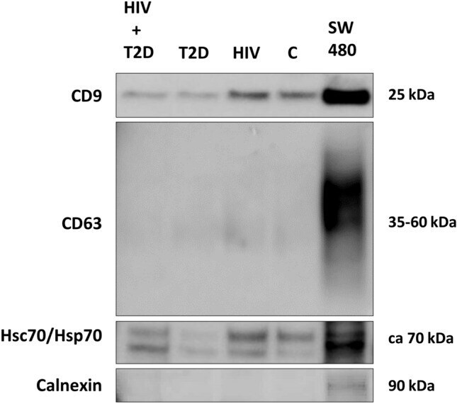
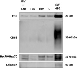
- Figure 2 Western blot detection of CD9, Hsc70/Hsp70 and Calnexin in plasma pool EVs. As a positive control, lysate from the colorectal cancer cell line SW480 was used. Full-length blots are presented in Supplementary Fig. 3 (Supplementary Information).