Explore
 Validate
 Learn
Learn Western blot
Antibody data
- Antibody Data
- Antigen structure
- References [3]
- Comments [0]
- Validations
- Western blot [3]
- Immunocytochemistry [1]
Submit
Validation data
Reference
Comment
Report error
- Product number
- MA5-12267 - Provider product page

- Provider
- Invitrogen Antibodies
- Product name
- TID1 Monoclonal Antibody (RS13)
- Antibody type
- Monoclonal
- Antigen
- Recombinant full-length protein
- Description
- MA5-12267 targets TID-1 in IP, IP, and WB applications and shows reactivity with Human, mouse, and Rat samples.
- Antibody clone number
- RS13
- Concentration
- 0.2 mg/mL
Submitted references p53Ψ is a transcriptionally inactive p53 isoform able to reprogram cells toward a metastatic-like state.
Tid1, CHIP and ErbB2 interactions and their prognostic implications for breast cancer patients.
Functional genetic screen for genes involved in senescence: role of Tid1, a homologue of the Drosophila tumor suppressor l(2)tid, in senescence and cell survival.
Senturk S, Yao Z, Camiolo M, Stiles B, Rathod T, Walsh AM, Nemajerova A, Lazzara MJ, Altorki NK, Krainer A, Moll UM, Lowe SW, Cartegni L, Sordella R
Proceedings of the National Academy of Sciences of the United States of America 2014 Aug 12;111(32):E3287-96
Proceedings of the National Academy of Sciences of the United States of America 2014 Aug 12;111(32):E3287-96
Tid1, CHIP and ErbB2 interactions and their prognostic implications for breast cancer patients.
Jan CI, Yu CC, Hung MC, Harn HJ, Nieh S, Lee HS, Lou MA, Wu YC, Chen CY, Huang CY, Chen FN, Lo JF
The Journal of pathology 2011 Nov;225(3):424-37
The Journal of pathology 2011 Nov;225(3):424-37
Functional genetic screen for genes involved in senescence: role of Tid1, a homologue of the Drosophila tumor suppressor l(2)tid, in senescence and cell survival.
Tarunina M, Alger L, Chu G, Munger K, Gudkov A, Jat PS
Molecular and cellular biology 2004 Dec;24(24):10792-801
Molecular and cellular biology 2004 Dec;24(24):10792-801
No comments: Submit comment
Supportive validation
- Submitted by
- Invitrogen Antibodies (provider)
- Main image
- Experimental details
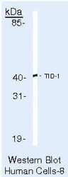
- Western blot of TID-1 using TID-1 Monoclonal Antibody (Product # MA5-12267) on HeLa Cells.
- Submitted by
- Invitrogen Antibodies (provider)
- Main image
- Experimental details
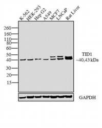
- Western blot analysis was performed on membrane enriched extracts (30 µg lysate) of K-562 (Lane 1), HEK-293 (Lane 2), Hep G2 (Lane 3), A549 (Lane 4), MCF7 (Lane 5), LNCaP (Lane 6) and tissue enriched extracts (30 µg lysate) of Rat Liver (Lane 7).The blots were probed with TID1 Mouse monoclonal Antibody (Product # MA5-12267, 2 µg/mL) and detected by chemiluminescence using Goat anti-Mouse IgG (H+L) Superclonal™ Secondary Antibody, HRP conjugate (Product # A28177, 0.25 µg/mL, 1:4000 dilution). A 40 kDa band corresponding to TID1 was observed across the cell lines tested. Apart from desired band, a 43 kDa band was observed in MCF7 and LNCap cell line representing the splice variant of the target. Known quantity of protein samples were electrophoresed using Novex® NuPAGE® 4-12 % Bis-Tris gel (Product # NP0322BOX), XCell SureLock™ Electrophoresis System (Product # EI0002) and Novex® Sharp Pre-Stained Protein Standard (Product # LC5800). Resolved proteins were then transferred onto a nitrocellulose membrane with iBlot® 2 Dry Blotting System (Product # IB21001). The membrane was probed with the relevant primary and secondary Antibody following blocking with 5% skimmed milk. Chemiluminescent detection was performed using Pierce™ ECL Western Blotting Substrate (Product # 32106).

- Submitted by
- Invitrogen Antibodies (provider)
- Main image

- Experimental details
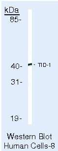
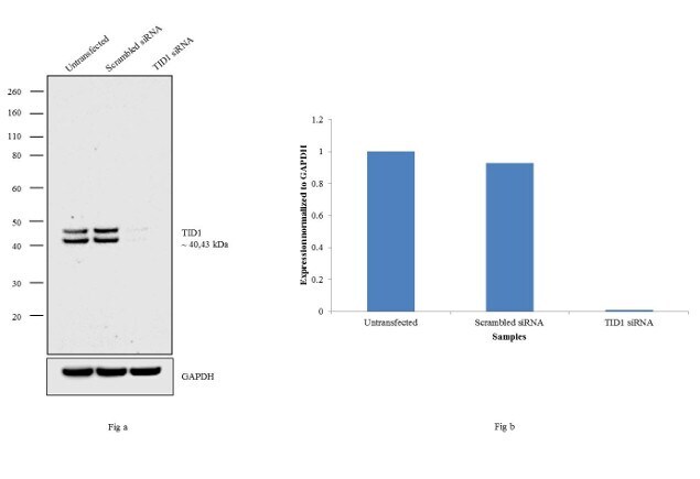
- Knockdown of TID1 was achieved by transfecting HeLa cells with TID1 specific siRNAs (Silencer® select Product # s17346, s17347). Western blot analysis (Fig a) was performed using whole cell extracts from the TID1 knock down cells (lane 3), non-specific scrambled siRNA transfected cells (lane 2) and untransfected cells (lane 1). The blots were probed with anti-TID1 Mouse monoclonal Antibody (Product # MA5-12267, 2 µg/mL) and Goat anti-Mouse IgG (H+L) Superclonal™ Secondary Antibody, HRP conjugate (Product # A28177, 0.25 µg/mL, 1:4000 dilution). Densitometric analysis of this western blot is shown in histogram (Fig b). Loss of signal upon siRNA mediated knock down confirms that antibody is specific to TID1.
Supportive validation
- Submitted by
- Invitrogen Antibodies (provider)
- Main image

- Experimental details
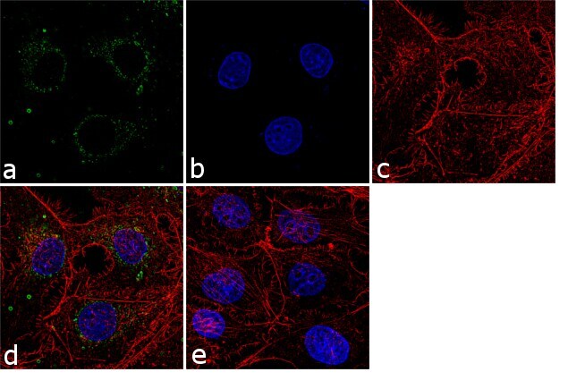
- Immunofluorescence analysis of TID1 was performed using 70% confluent log phase HepG2 cells. The cells were fixed with 4% paraformaldehyde for 10 minutes, permeabilized with 0.1% Triton™ X-100 for 10 minutes, and blocked with 1% BSA for 1 hour at room temperature. The cells were labeled with TID1 (RS13) Mouse Monoclonal Antibody (Product # MA5-12267) at 2 µg/mL in 0.1% BSA and incubated for 3 hours at room temperature and then labeled with Goat anti-Mouse IgG (H+L) Superclonal™ Secondary Antibody, Alexa Fluor® 488 conjugate (Product # A28175) at a dilution of 1:2000 for 45 minutes at room temperature (Panel a: green). Nuclei (Panel b: blue) were stained with SlowFade® Gold Antifade Mountant with DAPI (Product # S36938). F-actin (Panel c: red) was stained with Alexa Fluor® 555 Rhodamine Phalloidin (Product # R415, 1:300). Panel d represents the merged image showing cytoplasmic localization. Panel e shows the no primary antibody control. The images were captured at 60X magnification.