 Explore
 Validate
 Learn
Learn Western blot
 Immunohistochemistry
Antibody data
- Antibody Data
- Antigen structure
- References [1]
- Comments [0]
- Validations
- Western blot [3]
- Immunohistochemistry [6]
Submit
Validation data
Reference
Comment
Report error
- Product number
- HPA041656 - Provider product page

- Provider
- Atlas Antibodies
- Proper citation
- Atlas Antibodies Cat#HPA041656, RRID:AB_10960278
- Product name
- Anti-NUBP1
- Antibody type
- Polyclonal
- Reactivity
- Human
- Host
- Rabbit
- Conjugate
- Unconjugated
- Antigen sequence
TSDEHLSVVRYLATAHIDGAVIITTPQEVSLQDVR
KEINFCRKVKLPIIGVVEN- Isotype
- IgG
- Vial size
- 100 µl
- Storage
- Store at +4°C for short term storage. Long time storage is recommended at -20°C.
Submitted references The Diabetes Drug Target MitoNEET Governs a Novel Trafficking Pathway to Rebuild an Fe-S Cluster into Cytosolic Aconitase/Iron Regulatory Protein 1
Ferecatu I, Goncalves S, Golinelli-Cohen M, Clemancey M, Martelli A, Riquier S, Guittet E, Latour J, Puccio H, Drapier J, Lescop E, Bouton C
Journal of Biological Chemistry 2014 October;289(41):28070-28086
Journal of Biological Chemistry 2014 October;289(41):28070-28086
No comments: Submit comment
Supportive validation
Supportive validation
- Submitted by
- Atlas Antibodies (provider)
- Enhanced method
- Genetic validation
- Main image
- Experimental details
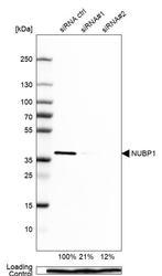
- Western blot analysis in MCF-7 cells transfected with control siRNA, target specific siRNA probe #1 and #2, using Anti-NUBP1 antibody. Remaining relative intensity is presented. Loading control: Anti-PPIB.

- Submitted by
- Atlas Antibodies (provider)
- Enhanced method
- Independent antibody validation
- Main image

- Experimental details
- Western blot analysis using Anti-NUBP1 antibody HPA041656 (A) shows similar pattern to independent antibody HPA041799 (B).

Supportive validation
- Submitted by
- Atlas Antibodies (provider)
- Main image

- Experimental details
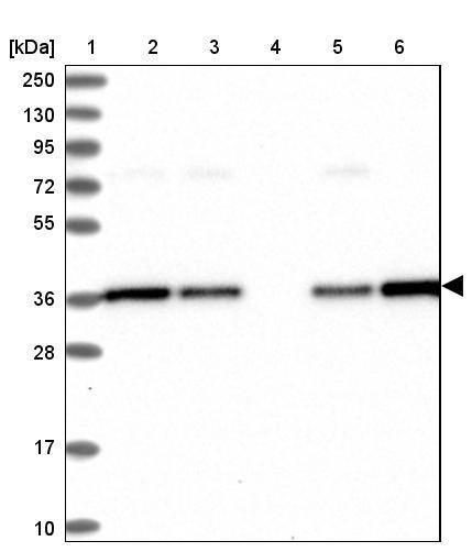
- Lane 1: Marker [kDa] 250, 130, 95, 72, 55, 36, 28, 17, 10Lane 2: Human cell line RT-4Lane 3: Human cell line U-251MG spLane 4: Human plasma (IgG/HSA depleted)Lane 5: Human liver tissueLane 6: Human tonsil tissue
Enhanced validation
Supportive validation
- Submitted by
- Atlas Antibodies (provider)
- Enhanced method
- Independent antibody validation
- Main image

- Experimental details
- Immunohistochemical staining of human colon, kidney, liver and testis using Anti-NUBP1 antibody HPA041656 (A) shows similar protein distribution across tissues to independent antibody HPA041799 (B).

Supportive validation
- Submitted by
- Atlas Antibodies (provider)
- Main image

- Experimental details
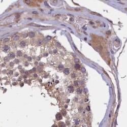
- Immunohistochemical staining of human testis shows moderate cytoplasmic positivity in cells in exocrine glandular cells.

- Submitted by
- Atlas Antibodies (provider)
- Main image
- Experimental details
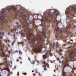
- Immunohistochemical staining of human colon using Anti-NUBP1 antibody HPA041656.
- Sample type
- HUMAN

- Submitted by
- Atlas Antibodies (provider)
- Main image

- Experimental details
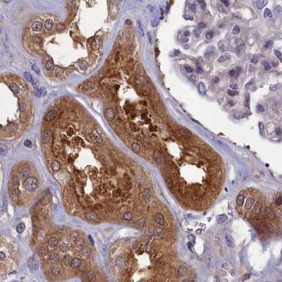
- Immunohistochemical staining of human kidney using Anti-NUBP1 antibody HPA041656.
- Sample type
- HUMAN
- Submitted by
- Atlas Antibodies (provider)
- Main image
- Experimental details
- Immunohistochemical staining of human testis using Anti-NUBP1 antibody HPA041656.
- Sample type
- HUMAN

- Submitted by
- Atlas Antibodies (provider)
- Main image
- Experimental details
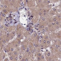
- Immunohistochemical staining of human liver using Anti-NUBP1 antibody HPA041656.
- Sample type
- HUMAN
