 Explore
 Validate
 Learn
Learn Western blot
 Other assay
Antibody data
- Antibody Data
- Antigen structure
- References [2]
- Comments [0]
- Validations
- Other assay [2]
Submit
Validation data
Reference
Comment
Report error
- Product number
- MA1-40244 - Provider product page

- Provider
- Invitrogen Antibodies
- Product name
- ASGR1 Monoclonal Antibody (8D7)
- Antibody type
- Monoclonal
- Antigen
- Other
- Description
- Applications Reported: This L243 antibody has been reported for use in flow cytometric analysis.
- Reactivity
- Human, Rat
- Host
- Mouse
- Isotype
- IgG
- Antibody clone number
- 8D7
- Vial size
- 100 µg
- Concentration
- 0.1 mg/mL
- Storage
- 4° C
Submitted references Generation of qualified clinical-grade functional hepatocytes from human embryonic stem cells in chemically defined conditions.
Maturation of induced pluripotent stem cell derived hepatocytes by 3D-culture.
Li Z, Wu J, Wang L, Han W, Yu J, Liu X, Wang Y, Zhang Y, Feng G, Li W, Stacey GN, Gu Q, Hu B, Wang L, Zhou Q, Hao J
Cell death & disease 2019 Oct 10;10(10):763
Cell death & disease 2019 Oct 10;10(10):763
Maturation of induced pluripotent stem cell derived hepatocytes by 3D-culture.
Gieseck RL 3rd, Hannan NR, Bort R, Hanley NA, Drake RA, Cameron GW, Wynn TA, Vallier L
PloS one 2014;9(1):e86372
PloS one 2014;9(1):e86372
No comments: Submit comment
Supportive validation
- Submitted by
- Invitrogen Antibodies (provider)
- Main image
- Experimental details
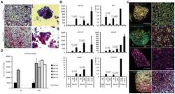
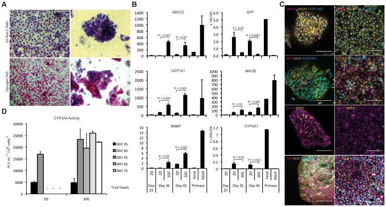
- Figure 2 Functional comparison of IPSC-Hep 3D clump culture versus traditional 2D culture. ( A ) Oil red O and periodic acid staining demonstrating lipid storage and glycogen synthesis in both 2D and 3D clump cultures. ( B ) qPCR analysis of select phase I and phase II enzymes, hepatic transporters, and other hepatic markers demonstrating a shift towards a more mature phenotype in the 3D clump cultures (fold expression to undifferentiated IPSCs; mean +- s.d.; n = 3 biological replicates). ( C ) Confocal micrographs comparing the presence and localization of hepatic markers within the two culture systems (scale bar = 100 microns). ( D ) CYP3A4 activity of the two culture conditions measured over a period of 75 days (mean +- s.d.; n = 3 biological replicates).
- Submitted by
- Invitrogen Antibodies (provider)
- Main image
- Experimental details
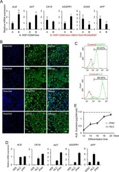
- Fig. 4 Differentiation of hESCs into HLCs. a The relative hepatocyte ( ALB , AAT , CK18 , and ASGPR1 ), cholangiocyte ( SOX9 ), and hepatoblast ( AFP ) gene expression levels of day 19 differentiated cells with different treatments (groups A and B) were determined by qPCR. HGF (hepatocyte growth factor, 20 ng/mL); OSM (oncostatin M, 20 ng/mL); Dex (dexamethasone, 10 uM); SB431542 (TGFbeta inhibitor, 2 uM); and RO4929097 (Notch inhibitor, 1 uM). b Immunofluorescence analysis of ALB, AAT, ASGPR1, and CK18 expression in group B-induced differentiated cells on day 19. c The expression levels of ALB and ASGPR1 in group B-induced differentiated cells were determined by flow cytometry on day 19. Isotype control antibodies were used as controls. d The relative hepatocyte ( ALB , AAT , CK18 , ASGPR1 , and AFP ) gene expression levels of differentiated HLCs (group B) compared with those of hESCs and primary human hepatocytes (PHHs) were determined by qPCR. e Albumin secretion of HLCs (black line) on days 12, 14, 16, 18, and 20 and PHHs (dotted line) on day 2 were determined by ELISA. * p < 0.05, ** p < 0.01; data are represented as the mean +- SD. Scale bar, 50 um.
