Explore
 Validate
 Learn
Learn Immunocytochemistry
 Immunohistochemistry
Antibody data
- Antibody Data
- Antigen structure
- References [0]
- Comments [0]
- Validations
- Immunocytochemistry [1]
- Immunohistochemistry [13]
Submit
Validation data
Reference
Comment
Report error
- Product number
- HPA003103 - Provider product page

- Provider
- Atlas Antibodies
- Proper citation
- Atlas Antibodies Cat#HPA003103, RRID:AB_10671088
- Product name
- Anti-USP11
- Antibody type
- Polyclonal
- Reactivity
- Human
- Host
- Rabbit
- Conjugate
- Unconjugated
- Antigen sequence
ESGRERPLRAGESWFLVEKHWYKQWEAYVQGGDQD
SSTFPGCINNATLFQDEINWRLKEGLVEGEDYVLL
PAAAWHYLVSWYGLEHGQPPIERKVIELPNIQKVE
VYPVELLLVRHNDLGKSHTVQFS- Isotype
- IgG
- Vial size
- 100 µl
- Storage
- Store at +4°C for short term storage. Long time storage is recommended at -20°C.
No comments: Submit comment
Supportive validation
- Submitted by
- Atlas Antibodies (provider)
- Main image
- Experimental details
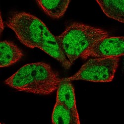
- Immunofluorescent staining of human cell line RH-30 shows localization to nucleoplasm & cytosol.
- Sample type
- HUMAN

Enhanced validation
Enhanced validation
Supportive validation
- Submitted by
- Atlas Antibodies (provider)
- Enhanced method
- Orthogonal validation
- Main image

- Experimental details
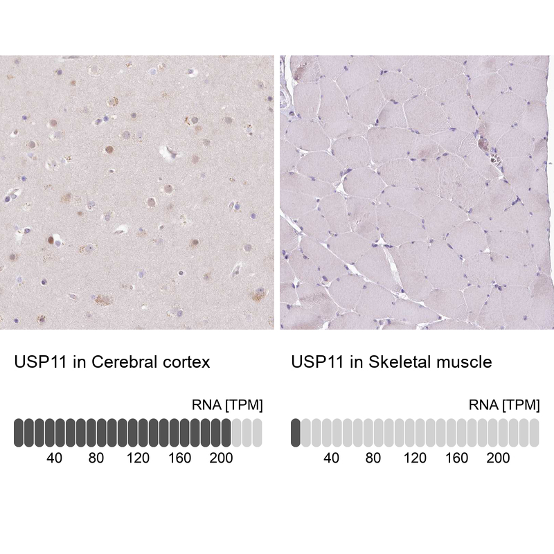
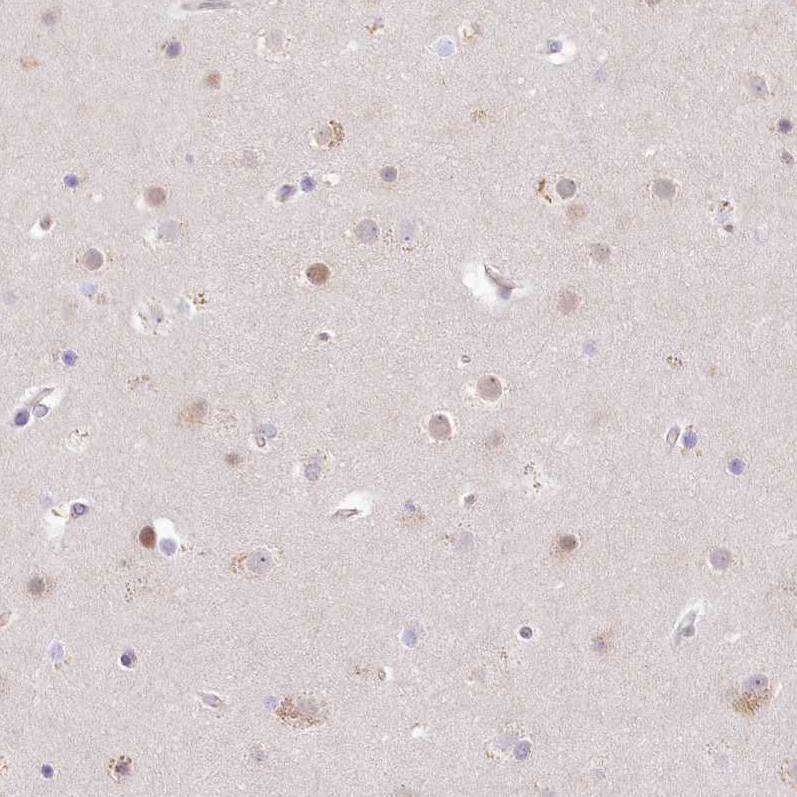
- Immunohistochemistry analysis in human cerebral cortex and skeletal muscle tissues using HPA003103 antibody. Corresponding USP11 RNA-seq data are presented for the same tissues.
- Sample type
- HUMAN
Enhanced validation
- Submitted by
- Atlas Antibodies (provider)
- Enhanced method
- Independent antibody validation
- Main image
- Experimental details
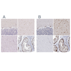
- Immunohistochemical staining of human cerebellum, cerebral cortex, skeletal muscle and testis using Anti-USP11 antibody HPA003103 (A) shows similar protein distribution across tissues to independent antibody HPA037536 (B).

Supportive validation
- Submitted by
- Atlas Antibodies (provider)
- Main image
- Experimental details
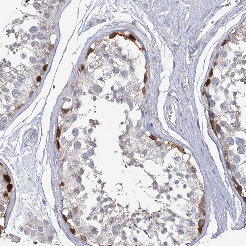
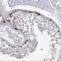
- Immunohistochemical staining of human testis shows distinct nuclear and cytoplasmic positivity of basal cells in seminiferus ducts.

- Submitted by
- Atlas Antibodies (provider)
- Main image

- Experimental details
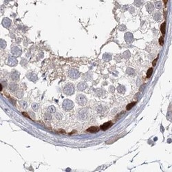
- Immunohistochemical staining of human testis shows high expression.
- Sample type
- HUMAN
- Submitted by
- Atlas Antibodies (provider)
- Main image

- Experimental details
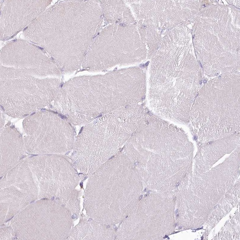
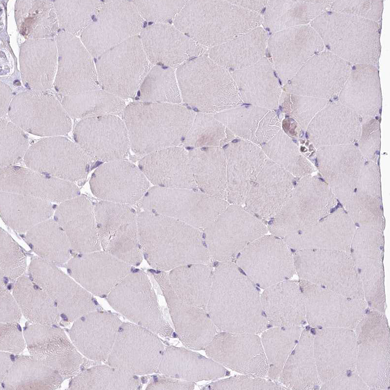
- Immunohistochemical staining of human skeletal muscle shows low expression as expected.
- Sample type
- HUMAN
- Submitted by
- Atlas Antibodies (provider)
- Main image
- Experimental details
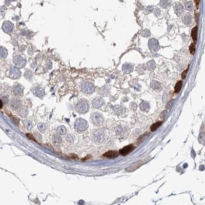
- Immunohistochemical staining of human testis using Anti-USP11 antibody HPA003103.
- Sample type
- HUMAN
- Submitted by
- Atlas Antibodies (provider)
- Main image
- Experimental details
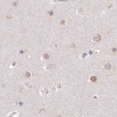
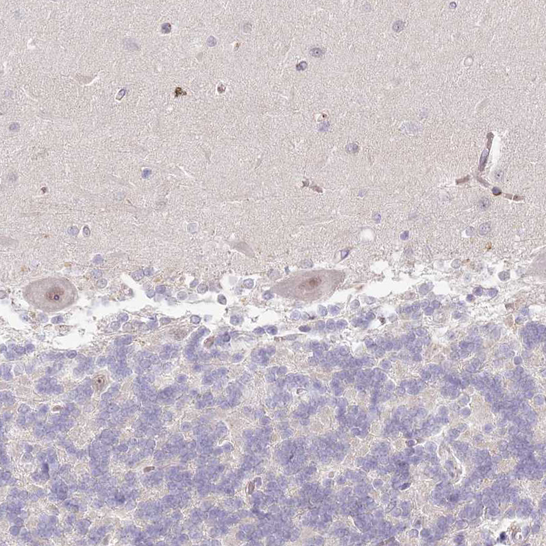
- Immunohistochemical staining of human cerebral cortex using Anti-USP11 antibody HPA003103.
- Sample type
- HUMAN
- Submitted by
- Atlas Antibodies (provider)
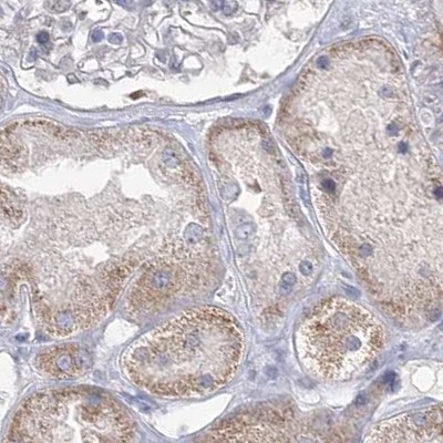
- Main image
- Experimental details
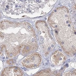
- Immunohistochemical staining of human kidney using Anti-USP11 antibody HPA003103.
- Sample type
- HUMAN
- Submitted by
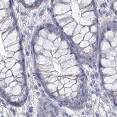
- Atlas Antibodies (provider)
- Main image
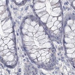
- Experimental details
- Immunohistochemical staining of human colon using Anti-USP11 antibody HPA003103.
- Sample type
- HUMAN
- Submitted by
- Atlas Antibodies (provider)
- Main image
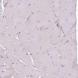
- Experimental details
- Immunohistochemical staining of human skeletal muscle shows no positivity in myocytes as expected.
- Sample type
- HUMAN
- Submitted by
- Atlas Antibodies (provider)
- Main image
- Experimental details
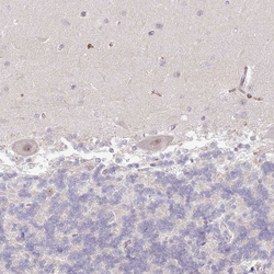
- Immunohistochemical staining of human cerebellum shows weak nuclear positivity in Purkinje cells.
- Sample type
- HUMAN
- Submitted by
- Atlas Antibodies (provider)
- Main image

- Experimental details
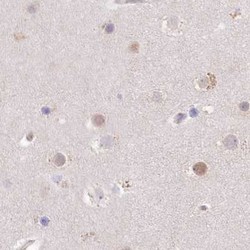
- Immunohistochemical staining of human cerebral cortex shows weak to moderate nuclear positivity in neurons.
- Sample type
- HUMAN
- Submitted by
- Atlas Antibodies (provider)
- Main image
- Experimental details
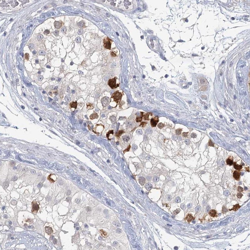
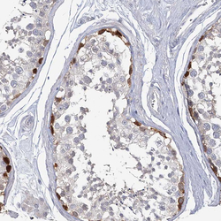
- Immunohistochemical staining of human testis shows strong nuclear positivity in a subset of cells in seminiferous ducts.
- Sample type
- HUMAN