Explore
 Validate
 Learn
Learn Western blot
 Immunocytochemistry
Antibody data
- Antibody Data
- Antigen structure
- References [1]
- Comments [0]
- Validations
- Western blot [1]
- Immunocytochemistry [1]
- Immunohistochemistry [11]
Submit
Validation data
Reference
Comment
Report error
- Product number
- HPA012123 - Provider product page

- Provider
- Atlas Antibodies
- Proper citation
- Atlas Antibodies Cat#HPA012123, RRID:AB_1849882
- Product name
- Anti-STIM1
- Antibody type
- Polyclonal
- Reactivity
- Human, Rat
- Host
- Rabbit
- Conjugate
- Unconjugated
- Antigen sequence
VHPGSLVEKLPDSPALAKKALLALNHGLDKAHSLM
ELSPSAPPGGSPHLDSSRSHSPSSPDPDTPSPVGD
SRALQASRNTRIPHLAGKKAVAEEDNGSIGEETDS
SPGRKKFPLKIFK- Isotype
- IgG
- Vial size
- 100 µl
- Storage
- Store at +4°C for short term storage. Long time storage is recommended at -20°C.
Submitted references STIM1 and SLC24A4 Are Critical for Enamel Maturation.
Wang S, Choi M, Richardson AS, Reid BM, Seymen F, Yildirim M, Tuna E, Gençay K, Simmer JP, Hu JC
Journal of dental research 2014 Jul;93(7 Suppl):94S-100S
Journal of dental research 2014 Jul;93(7 Suppl):94S-100S
No comments: Submit comment
Enhanced validation
- Submitted by
- Atlas Antibodies (provider)
- Enhanced method
- Orthogonal validation
- Main image
- Experimental details
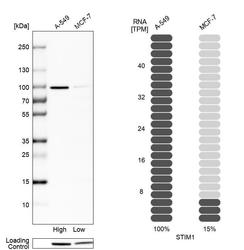
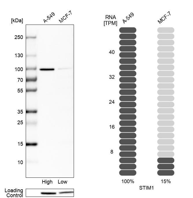
- Western blot analysis in human cell lines A-549 and MCF-7 using Anti-STIM1 antibody. Corresponding STIM1 RNA-seq data are presented for the same cell lines. Loading control: Anti-HSP90B1.
Supportive validation
- Submitted by
- Atlas Antibodies (provider)
- Main image
- Experimental details
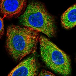
- Immunofluorescent staining of human cell line A549 shows localization to endoplasmic reticulum.
- Sample type
- HUMAN

Enhanced validation
Enhanced validation
Supportive validation
- Submitted by
- Atlas Antibodies (provider)
- Enhanced method
- Orthogonal validation
- Main image
- Experimental details
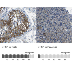
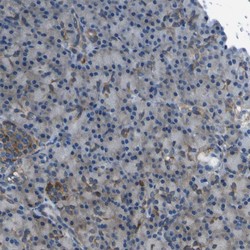
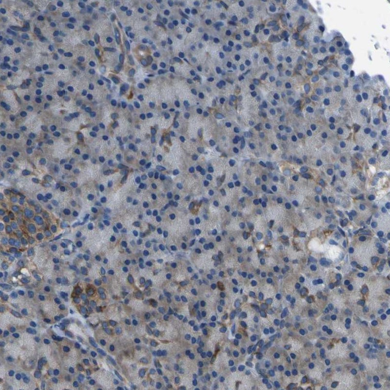
- Immunohistochemistry analysis in human testis and pancreas tissues using Anti-STIM1 antibody. Corresponding STIM1 RNA-seq data are presented for the same tissues.
- Sample type
- HUMAN

Enhanced validation
- Submitted by
- Atlas Antibodies (provider)
- Enhanced method
- Independent antibody validation
- Main image
- Experimental details
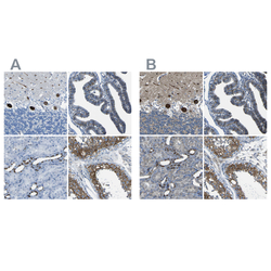
- Immunohistochemical staining of human cerebellum, fallopian tube, kidney and testis using Anti-STIM1 antibody HPA012123 (A) shows similar protein distribution across tissues to independent antibody HPA011088 (B).

Supportive validation
- Submitted by
- Atlas Antibodies (provider)
- Main image
- Experimental details
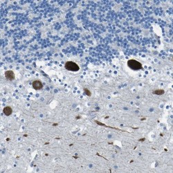
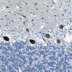
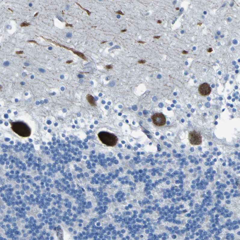
- Immunohistochemical staining of human cerebellum shows strong cytoplasmic positivity in Purkinje cells.

- Submitted by
- Atlas Antibodies (provider)
- Main image

- Experimental details
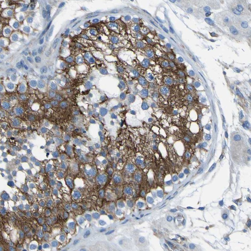
- Immunohistochemical staining of human testis shows high expression.
- Sample type
- HUMAN
- Submitted by
- Atlas Antibodies (provider)
- Main image
- Experimental details
- Immunohistochemical staining of human pancreas shows low expression as expected.
- Sample type
- HUMAN
- Submitted by
- Atlas Antibodies (provider)
- Main image
- Experimental details
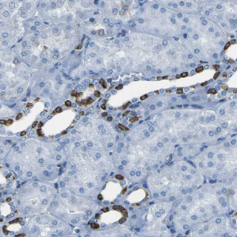
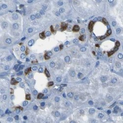
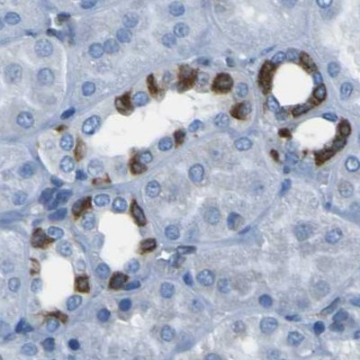
- Immunohistochemical staining of human kidney using Anti-STIM1 antibody HPA012123.
- Sample type
- HUMAN
- Submitted by
- Atlas Antibodies (provider)
- Main image
- Experimental details
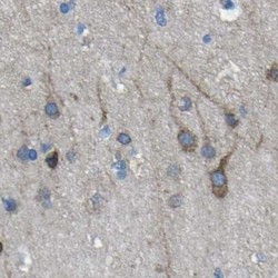
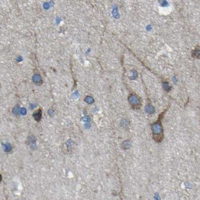
- Immunohistochemical staining of human cerebral cortex using Anti-STIM1 antibody HPA012123.
- Sample type
- HUMAN
- Submitted by
- Atlas Antibodies (provider)
- Main image
- Experimental details
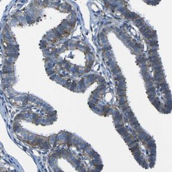
- Immunohistochemical staining of human fallopian tube shows strong cytoplasmic positivity in glandular cells.
- Sample type
- HUMAN

- Submitted by
- Atlas Antibodies (provider)
- Main image
- Experimental details
- Immunohistochemical staining of human cerebellum shows strong cytoplasmic positivity in Purkinje cells.
- Sample type
- HUMAN
- Submitted by
- Atlas Antibodies (provider)
- Main image
- Experimental details
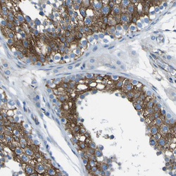
- Immunohistochemical staining of human testis shows strong cytoplasmic positivity in cells in seminiferous ducts.
- Sample type
- HUMAN

- Submitted by
- Atlas Antibodies (provider)
- Main image
- Experimental details
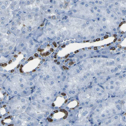
- Immunohistochemical staining of human kidney shows strong cytoplasmic positivity in cells in tubules.
- Sample type
- HUMAN