Explore
 Validate
 Learn
Learn Western blot
 Immunocytochemistry
Antibody data
- Antibody Data
- Antigen structure
- References [0]
- Comments [0]
- Validations
- Western blot [3]
- Immunocytochemistry [5]
- Immunohistochemistry [13]
Submit
Validation data
Reference
Comment
Report error
- Product number
- AMAb90662 - Provider product page

- Provider
- Atlas Antibodies
- Proper citation
- Atlas Antibodies Cat#AMAb90662, RRID:AB_2665623
- Product name
- Anti-ANLN
- Antibody type
- Monoclonal
- Reactivity
- Human
- Host
- Mouse
- Conjugate
- Unconjugated
- Antigen sequence
IVKSTLSQTVPSKGELSREICLQSQSKDKSTTPGG
TGIKPFLERFGERCQEHSKESPARSTPHRTPIITP
NTKAIQERLFKQDTSSSTTHLAQQLKQERQKELAC
LRGRFDKGNIWSAEKGGNSKSKQLETKQETH- Epitope
- Binds to an epitope located within the peptide sequence LRGRFDKGNIWSAEK as determined by overlapping synthetic peptides.
- Isotype
- IgG
- Antibody clone number
- CL0303
- Vial size
- 100 µl
- Storage
- Store at +4°C for short term storage. Long time storage is recommended at -20°C.
No comments: Submit comment
Enhanced validation
- Submitted by
- Atlas Antibodies (provider)
- Enhanced method
- Genetic validation
- Main image
- Experimental details
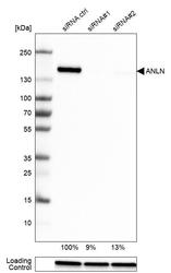
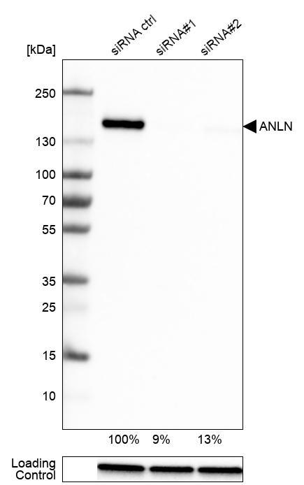
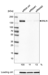
- Western blot analysis in U-251MG cells transfected with control siRNA, target specific siRNA probe #1 and #2, using Anti-ANLN antibody. Remaining relative intensity is presented. Loading control: Anti-GAPDH.
- Submitted by
- Atlas Antibodies (provider)
- Main image
- Experimental details
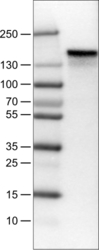
- Lane 1: Marker [kDa]Lane 2:Human cell line U-251 MG

- Submitted by
- Atlas Antibodies (provider)
- Main image
- Experimental details
- Western blot analysis of extracts from U-251 cells, transfected with: control siRNA, target specific siRNA probe #1, target specific siRNA probe #2, using Anti-ANLN monoclonal antibody. Downregulation of antibody signal confirms target specificity. Remaining % intensity, relative control lane, is indicated. Anti-GAPDH monoclonal antibody was used as loading control.

Supportive validation
- Submitted by
- Atlas Antibodies (provider)
- Main image
- Experimental details
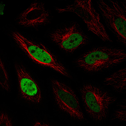
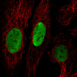
- Immunofluorescence staining in HeLa cell line with Anti-ANLN monoclonal antibody, showing cell cycle dependent nuclear (without nucleoli) staining in green. Microtubule- and nuclear probes are visualized in red and blue respectively (where available).
- Sample type
- HUMAN

- Submitted by
- Atlas Antibodies (provider)
- Main image
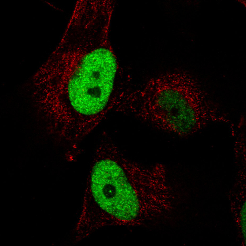
- Experimental details
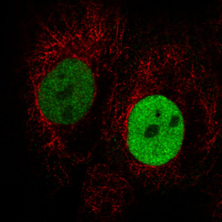
- Immunofluorescence staining in A431 cell line with Anti-ANLN monoclonal antibody, showing cell cycle dependent nuclear (without nucleoli) staining in green. Microtubule- and nuclear probes are visualized in red and blue respectively (where available).
- Sample type
- HUMAN

- Submitted by
- Atlas Antibodies (provider)
- Main image

- Experimental details
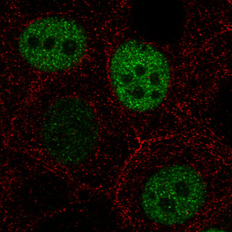
- Immunofluorescence staining in MCF7 cell line with Anti-ANLN monoclonal antibody, showing cell cycle dependent nuclear (without nucleoli) staining in green. Microtubule- and nuclear probes are visualized in red and blue respectively (where available).
- Sample type
- HUMAN
- Submitted by
- Atlas Antibodies (provider)
- Main image
- Experimental details
- Immunofluorescence staining in U2OS cell line with Anti-ANLN monoclonal antibody, showing cell cycle dependent nuclear (without nucleoli) staining in green. Microtubule- and nuclear probes are visualized in red and blue respectively (where available).
- Sample type
- HUMAN

- Submitted by
- Atlas Antibodies (provider)
- Main image

- Experimental details
- Immunofluorescence staining in U251 cell line with Anti-ANLN monoclonal antibody, showing cell cycle dependent nuclear (without nucleoli) staining in green. Microtubule- and nuclear probes are visualized in red and blue respectively (where available).
- Sample type
- HUMAN
Enhanced validation
Supportive validation
- Submitted by
- Atlas Antibodies (provider)
- Enhanced method
- Orthogonal validation
- Main image
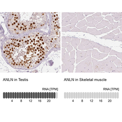
- Experimental details
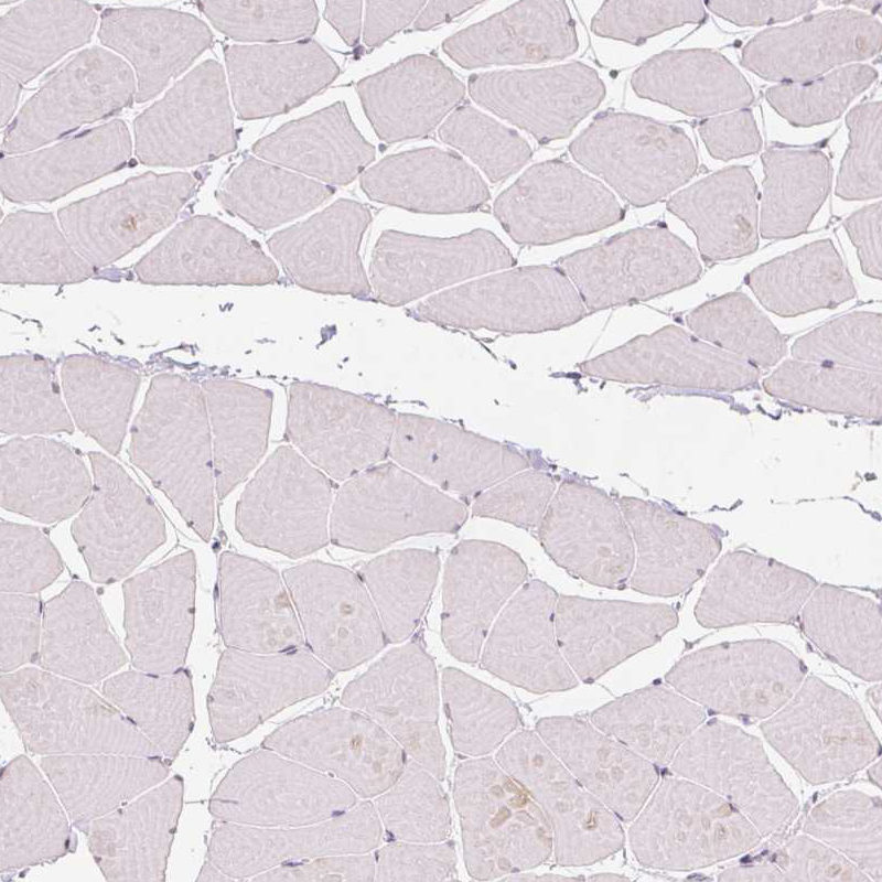
- Immunohistochemistry analysis in human testis and skeletal muscle tissues using AMAb90662 antibody. Corresponding ANLN RNA-seq data are presented for the same tissues.
- Sample type
- HUMAN

Supportive validation
- Submitted by
- Atlas Antibodies (provider)
- Main image
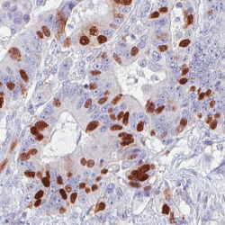
- Experimental details
- Immunohistochemical staining of human colorectal cancer shows strong nuclear immunoreactivity in a subset of tumor cells.

- Submitted by
- Atlas Antibodies (provider)
- Main image

- Experimental details
- Immunohistochemical staining of human lung cancer shows strong nuclear positivity in a subset of tumor cells.
- Submitted by
- Atlas Antibodies (provider)
- Main image
- Experimental details
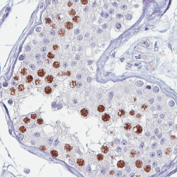
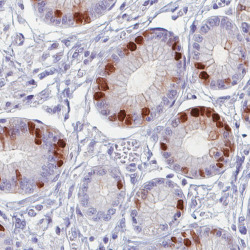
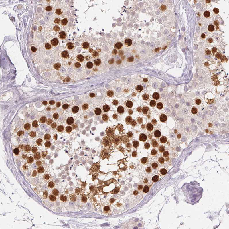
- Immunohistochemical staining of human testis shows strong immunoreactivity in a subset of cells in seminiferous tubules.
- Submitted by
- Atlas Antibodies (provider)
- Main image
- Experimental details
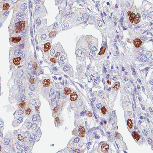
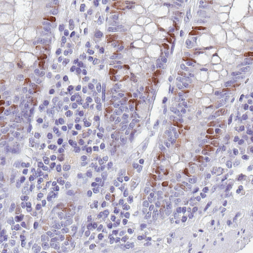
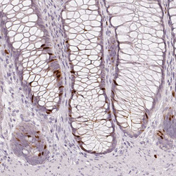
- Immunohistochemical staining of human colon shows strong positivity in some glandular cells.
- Submitted by
- Atlas Antibodies (provider)
- Main image
- Experimental details
- Immunohistochemical staining of human stomach shows strong nuclear positivity in glandular cells.

- Submitted by
- Atlas Antibodies (provider)
- Main image

- Experimental details
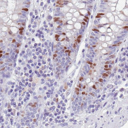
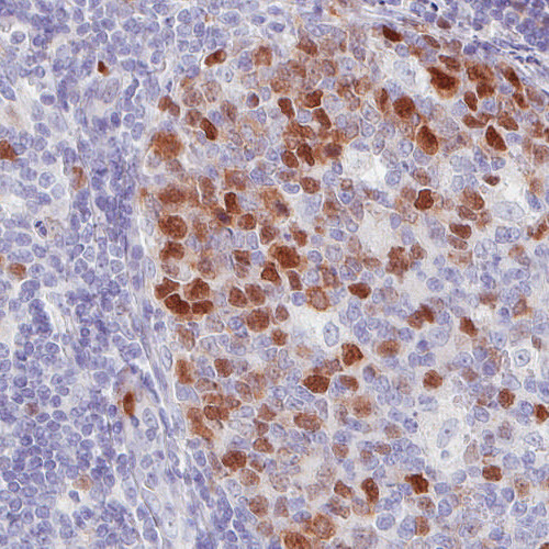
- Immunohistochemical staining of human tonsil shows strong immunoreactivity in the germinal center lymphoid cells.
- Submitted by
- Atlas Antibodies (provider)
- Main image

- Experimental details
- Immunohistochemical staining of human liver shows absence of staining (negative control).

- Submitted by
- Atlas Antibodies (provider)
- Main image
- Experimental details
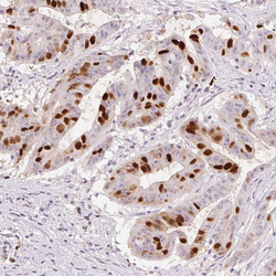
- Immunohistochemical staining of human colorectal cancer shows moderate to strong nuclear positivity in a subset of tumor cells.

- Submitted by
- Atlas Antibodies (provider)
- Main image

- Experimental details
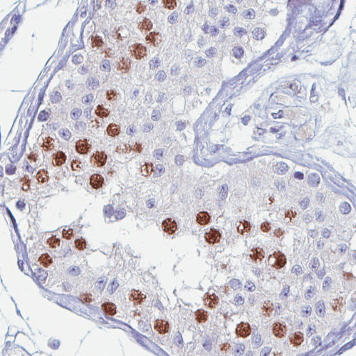
- Immunohistochemical staining of human testis shows moderate to strong nuclear positivity in a subset of cells in seminiferous ducts.
- Submitted by
- Atlas Antibodies (provider)
- Main image
- Experimental details
- Immunohistochemical staining of human rectum shows moderate to strong nuclear positivity in a subset of glandular cells.

- Submitted by
- Atlas Antibodies (provider)
- Main image
- Experimental details
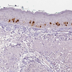
- Immunohistochemical staining of human skin shows moderate to strong nuclear positivity in a subset of dermal cells.

- Submitted by
- Atlas Antibodies (provider)
- Main image
- Experimental details
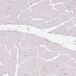
- Immunohistochemical staining of human skeletal muscle shows no nuclear positivity in striated muscle fibers as expected.