 Explore
 Validate
 Learn
Learn Western blot
Antibody data
- Antibody Data
- Antigen structure
- References [8]
- Comments [0]
- Validations
- Western blot [2]
- Immunocytochemistry [1]
- Other assay [8]
Submit
Validation data
Reference
Comment
Report error
- Product number
- PA5-19810 - Provider product page

- Provider
- Invitrogen Antibodies
- Product name
- TNF alpha Polyclonal Antibody
- Antibody type
- Polyclonal
- Antigen
- Synthetic peptide
- Description
- This antibody is predicted to react with rat, cat, chimpanzee and baboon based on sequence homology.
- Concentration
- 1 mg/mL
Submitted references Design of Targeted Flurbiprofen Biomimetic Nanoparticles for Management of Arthritis: In Vitro and In Vivo Appraisal.
Successful Treatment of Noise-Induced Hearing Loss by Mesenchymal Stromal Cells: An RNAseq Analysis of Protective/Repair Pathways.
Oxidation of Hemoglobin Drives a Proatherogenic Polarization of Macrophages in Human Atherosclerosis.
Improved diabetic wound healing by LFcinB is associated with relevant changes in the skin immune response and microbiota.
Hydrogen sulfide inhibits aortic valve calcification in heart via regulating RUNX2 by NF-κB, a link between inflammation and mineralization.
Protective effect of mild-induced hypothermia against moderate traumatic brain injury in rats involved in necroptotic and apoptotic pathways.
Anti-tumor effects of Abnormal Savda Munziq on the transplanted cervical cancer (U27) mouse model.
Fracture induces keratinocyte activation, proliferation, and expression of pro-nociceptive inflammatory mediators.
Mohamed HI, El-Kamel AH, Hammad GO, Heikal LA
Pharmaceutics 2022 Jan 7;14(1)
Pharmaceutics 2022 Jan 7;14(1)
Successful Treatment of Noise-Induced Hearing Loss by Mesenchymal Stromal Cells: An RNAseq Analysis of Protective/Repair Pathways.
Warnecke A, Harre J, Shew M, Mellott AJ, Majewski I, Durisin M, Staecker H
Frontiers in cellular neuroscience 2021;15:656930
Frontiers in cellular neuroscience 2021;15:656930
Oxidation of Hemoglobin Drives a Proatherogenic Polarization of Macrophages in Human Atherosclerosis.
Potor L, Hendrik Z, Patsalos A, Katona É, Méhes G, Póliska S, Csősz É, Kalló G, Komáromi I, Combi Z, Posta N, Sikura KÉ, Pethő D, Oros M, Vereb G, Tóth C, Gergely P, Nagy L, Balla G, Balla J
Antioxidants & redox signaling 2021 Oct 20;35(12):917-950
Antioxidants & redox signaling 2021 Oct 20;35(12):917-950
Improved diabetic wound healing by LFcinB is associated with relevant changes in the skin immune response and microbiota.
Mouritzen MV, Petkovic M, Qvist K, Poulsen SS, Alarico S, Leal EC, Dalgaard LT, Empadinhas N, Carvalho E, Jenssen H
Molecular therapy. Methods & clinical development 2021 Mar 12;20:726-739
Molecular therapy. Methods & clinical development 2021 Mar 12;20:726-739
Hydrogen sulfide inhibits aortic valve calcification in heart via regulating RUNX2 by NF-κB, a link between inflammation and mineralization.
Éva Sikura K, Combi Z, Potor L, Szerafin T, Hendrik Z, Méhes G, Gergely P, Whiteman M, Beke L, Fürtös I, Balla G, Balla J
Journal of advanced research 2021 Jan;27:165-176
Journal of advanced research 2021 Jan;27:165-176
Protective effect of mild-induced hypothermia against moderate traumatic brain injury in rats involved in necroptotic and apoptotic pathways.
Zhang HB, Cheng SX, Tu Y, Zhang S, Hou SK, Yang Z
Brain injury 2017;31(3):406-415
Brain injury 2017;31(3):406-415
Anti-tumor effects of Abnormal Savda Munziq on the transplanted cervical cancer (U27) mouse model.
Omarniyaz Z, Yu Y, Yang T, Shan L, Miao W, Reyimu R, Upur H, Aikemu A
BMC complementary and alternative medicine 2016 Nov 24;16(1):477
BMC complementary and alternative medicine 2016 Nov 24;16(1):477
Fracture induces keratinocyte activation, proliferation, and expression of pro-nociceptive inflammatory mediators.
Li WW, Guo TZ, Li XQ, Kingery WS, Clark DJ
Pain 2010 Dec;151(3):843-852
Pain 2010 Dec;151(3):843-852
No comments: Submit comment
Supportive validation
- Submitted by
- Invitrogen Antibodies (provider)
- Main image

- Experimental details
- Western blot analysis of HeLa Whole Cell Lysate using Product # PA5-19810, TNF alpha primary antibody at a dilution of 1 µg/mL (lane 1). Staining of Mouse Liver Tissue Lysate at a dilution of 1 µg/mL (lane 2). Blot treated with a secondary HRP-conjugated Goat polyclonal anti-Rabbit antibody was used at a dilution of 1:3000.

- Submitted by
- Invitrogen Antibodies (provider)
- Main image

- Experimental details
- Western blot was performed using Anti-TNF alpha Polyclonal Antibody (Product # PA5-19810) and a 25kDa band corresponding to TNF-Alpha was detected in lipopolysaccharide treated macrophages (differentiated from both THP-1 and U-937 and RAW 264.7 macrophage). Whole cell extracts (30 µg lysate) of untreated macrophages differentiated from U-937 (Lane 1), treated with PTI (Lane 2), stimulated with LPS (Lane 3), stimulated with LPS and subsequently treated with PTI (Lane 4), untreated RAW 264.7 (Lane 5), stimulated with LPS (Lane 6), stimulated with LPS and subsequently treated with PTI (Lane 7) in Fig. a, and untreated, differentiated macrophage from THP-1 (Lane 1), differentiated macrophage treated with PTI (Lane 2), THP-1 macrophage stimulated with LPS (Lane 3), THP-1 macrophage stimulated with LPS and then treated with PTI (Lane 4) in Fig. b were electrophoresed using NuPAGE™ 12% Bis-Tris Protein Gel (Product # NP0341BOX). Resolved proteins were then transferred onto a nitrocellulose membrane (Product # IB23001) by iBlot® 2 Dry Blotting System (Product # IB21001). The blot was probed with the primary antibody (0.9 µg/mL) and detected by chemiluminescence with Goat anti-Rabbit IgG (H+L) Superclonal™ Recombinant Secondary Antibody, HRP (Product # A27036, 1:8000 dilution) using the iBright FL 1000 (Product # A32752). Chemiluminescent detection was performed using Novex® ECL Reagent Kit (Product # WP20005).

Supportive validation
- Submitted by
- Invitrogen Antibodies (provider)
- Main image

- Experimental details
- Immunofluorescence analysis of Tumor necrosis factor alpha was performed using 80% confluent log phase RAW 264.7 treated with lipopolysaccharide (LPS) (10 ng/mL for 7 hr) and protein transport inhibitor cocktail [500X] (PTI) (1X for 4 hr). The cells were fixed with 4% paraformaldehyde for 10 minutes, permeabilized with 0.1% Triton™ X-100 for 10 minutes, and blocked with 2% BSA for 45 minutes at room temperature. The cells were labeled with TNF alpha Polyclonal Antibody (Product # PA5-19810) at 1 µg/mL in 0.1% BSA, incubated at 4 degree celsius overnight and then labeled with Donkey anti-Rabbit IgG (H+L) Highly Cross-Adsorbed Secondary Antibody, Alexa Fluor Plus 488 (Product # A32790), (1:2500), for 45 minutes at room temperature (Panel a,e,i: Green). Nuclei (Panel b,f,j: Blue) were stained with ProLong™ Diamond Antifade Mountant with DAPI (Product # P36962). F-actin (Panel c,g,k: Red) was stained with Rhodamine Phalloidin (Product # R415, 1:300). Panel d represents the merged image of untreated cells showing no staining for TNF-alpha and panel h and i shows upregulation of TNF-alpha expression in golgi-like structures upon LPS stimulation only and intracellular signal accumulation upon subsequent treatment with the secretory blocker PTI, respectively. The images were captured at 60X magnification.

Supportive validation
- Submitted by
- Invitrogen Antibodies (provider)
- Main image

- Experimental details
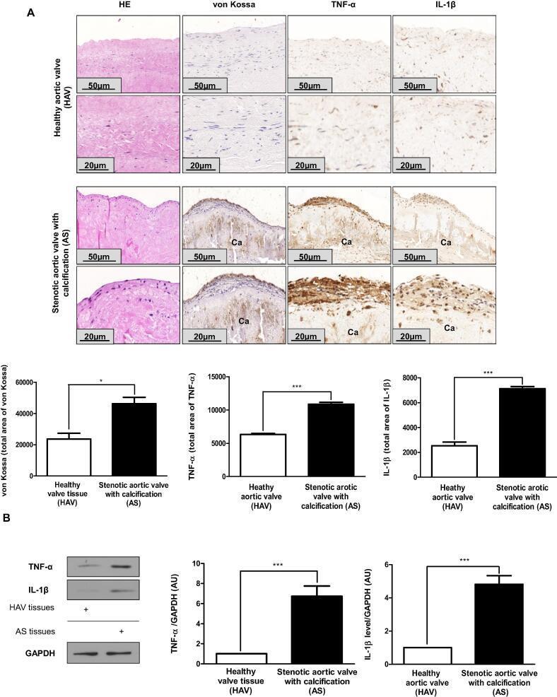
- Fig. 1 Inflammatory cytokines are characteristic of human stenotic aortic valve. A) Hematoxylin and eosin, von Kossa, TNF-alpha and IL1-beta IHC staining were performed on HAV valves derived from the Department of Forensic Institute, University of Debrecen (upper images; N = 3) and on AS valves (lower images, N = 5). Scale bars (50 um and 20 um) and pixel intensity of IHC staining were shown. Representative staining was shown. B) IL1-beta and TNF-alpha protein expression in HAV and AS heart valve tissue was assessed by western blot analysis. Protein expressions were normalized to GAPDH. Results were analyzed by One Way ANOVA, Bonferroni's Multiple Comparison Test and were shown as mean values +- SEM of five independent experiments. *P < 0.01; ***P < 0.0001.
- Submitted by
- Invitrogen Antibodies (provider)
- Main image

- Experimental details
- Fig. 2 Hydrogen sulfide inhibits inflammation and calcification. A) TNF-alpha and IL1-beta staining were performed on aortic valves of ApoE -/- mice kept on high-fat diet (upper panels, N = 5) and on high-fat diet treated with AP72 (lower panels; N = 5). B) Alizarin Red S staining was performed in ex vivo cultured aortic valves of ApoE -/- mice cultured in growth medium or calcification medium in the absence or presence with AP72 were shown. Results were analyzed by One Way ANOVA, Bonferroni's Multiple Comparison Test and were shown as mean values +- SEM of five independent experiments. **P < 0.001; ***P < 0.0001.

- Submitted by
- Invitrogen Antibodies (provider)
- Main image

- Experimental details
- Fig. 3 Hydrogen sulfide controls pro-inflammatory cytokines expression. A) IL1-beta, B) TNF-alpha western blot and RT-qPCR were performed and normalized to GAPDH. C) VIC cultured in growth medium or calcification medium in the absence or presence with AP72. Representative Alizarin Red S staining of human AS derived VIC was shown. VIC were cultured in growth medium or calcification medium. Western blots were carrying out from CSE and CBS double gene silencing utilizing siRNA. D) IL1-beta and E) TNF-alpha western blot was showing. Western blots were normalized to GAPDH. Results were analyzed by One Way ANOVA, Bonferroni's Multiple Comparison Test and were shown as mean values +- SEM of five independent experiments. *P < 0.01; **P < 0.001; ***P < 0.0001.

- Submitted by
- Invitrogen Antibodies (provider)
- Main image

- Experimental details
- Figure 5 Immunofluorescence analysis of macrophage abundance and phenotype and production of reactive oxygen species (ROS) in diabetic and non-diabetic mouse skin treated with LFcinB Diabetic mice (left) or healthy non-diabetic mice (right) were treated either with saline or with a low (12.5 mug/wound) or high (25 mug/wound) dose of LFcinB for 10 days. (A and B) Representative confocal microscopy images of CD68/TNF-alpha (M1 macrophages) and CD68/CD206 (M2 macrophages) positive stained cells in diabetic and healthy mouse skin sections. (C and D) Representative images of ROS production in murine wound skin measured by dihydroethidium (DHE) staining in diabetic or healthy animals. (E-H) The number of macrophages was quantified as the average number of CD68 positive cells co-stained with TNF-alpha for M1 and CD206 for M2 macrophages. (I and K) Pro-inflammatory/anti-inflammatory phenotype evaluated as M1/M2 ratio for diabetic or healthy mice. (J and L) ROS were measured as integrated density gray value after detection of the red signal of DHE staining in diabetic or healthy skin sections. Magnifications, 400x for CD68/TNF-alpha and CD68/CD206 and 200x for DHE. Scale bars, 100 mum for CD68/TNF-alpha and CD68/CD206 and 200 mum for DHE. The graph represents mean +- SD of an experiment (n = 4 animals per group), comparing the difference between the saline and treatment groups or between treatment groups (*) and the comparison of saline to baseline (#).* ,# p < 0.05, **p < 0.01, ***p

- Submitted by
- Invitrogen Antibodies (provider)
- Main image

- Experimental details
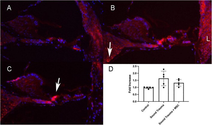
- FIGURE 7 Expression of TNFalpha protein (Red A-C ) and semi quantitative RT PCR of TNFalpha mRNA (D) . Control cochlea showed minimal TNF expression in the hair cells and lateral wall (A) . After sound trauma there was a clear increase in TFN alpha immunolabeling in hair cells, the spiral ganglion (arrow) and the lateral wall (B) . Treatment with MSCs after sound trauma reduced TNFalpha expression in the lateral wall and outer hair cells but expression remained high in the inner hair cell (arrow C ). Semi quantitative RT-PCR showed an up-regulation of TNF gene expression after sound trauma. This was slightly reduced after MSC treatment but not to control levels (D) .
- Submitted by
- Invitrogen Antibodies (provider)
- Main image

- Experimental details
- Figure 8 Western blot demonstration of TNF-alpha and IL-6 expression patterns in the articular cartilages of groups (A-F) 2-week post-treatment. where A is the healthy group, B is the untreated arthritic group, C is the group treated with FLUR-loaded HA-BSA NPs, D is the group treated with blank HA-BSA NPs, E is the group treated with FLUR-loaded BSA NPs and F is the group treated with blank BSA NPs. ( a ) Representative image of western blot showing the expression pattern of IL-6, TNF- alpha and beta-actin, ( b ) Normalized IL-6 expression in all group samples against beta-actin and ( c ) Normalized TNF-alpha expression in all group samples against beta-actin. Statistical significance is shown where **** p

- Submitted by
- Invitrogen Antibodies (provider)
- Main image

- Experimental details
- Figure 5. Effects of HT on TNF- a , TRAIL, FasL, FADD, caspase 3, caspase 8, PARP-1 and RIPK-3 protein expressions after moderate TBI in rats. TNF- a , TRAIL, FasL, FADD, caspase 3, caspase 8, PARP-1 and RIPK-3 protein expressions were determined by western blot. b -actin was used as an inner control. Data are shown as mean +- SD ( n = 5/group). * p < 0.05, ** p < 0.01 vs sham+NT; # p < 0.05 TBI+HT vs TBI+NT.

- Submitted by
- Invitrogen Antibodies (provider)
- Main image

- Experimental details
- FIG. 6. Ferryl hemoglobin-positive macrophages in carotid artery are positive for IL-1beta and TNF-alpha. Cross sections of a healthy carotid artery and atheromatous and hemorrhagic transformed lesions were shown. (A) Images demonstrated macrophages positive for both CD68 and IL-1beta in the atheromatous plaque and positive for ferrylHb, CD68, and IL-1beta in the hemorrhagic transformed lesions. Sections were stained with Hoechst 33258 for DNA ( blue ), an anti-ferrylHb antibody with Alexa Flour 488 secondary antibody for ferrylHb ( green ), an anti-CD68 antibody with Alexa Flour 568 secondary antibody for CD68 ( yellow ), and an anti-IL-1beta antibody with Alexa Flour 647 secondary antibody for IL-1beta ( red ). (B) Images demonstrated macrophages positive for both CD68 and TNF-alpha in the atheromatous plaque and positive for ferrylHb, CD68, and TNF-alpha in the hemorrhagic transformed lesions. Sections were stained with Hoechst 33258 for DNA ( blue ), an anti-ferrylHb antibody with Alexa Flour 488 secondary antibody for ferrylHb ( green ), an anti-CD68 antibody with Alexa Flour 568 secondary antibody for CD68 ( yellow ), and an anti-TNF-alpha antibody with Alexa Flour 647 secondary antibody for TNF-alpha ( red ). Images were taken using Leica TCS SP8 gated STED-CW nanoscopy. Images were deconvolved using Huygens Professional software. Representative image, N = 5. (C) Fluorescence intensity for ferrylHb ( n = 17), CD68 ( n = 20), IL-1beta ( n = 19), and TNF-alpha ( n = 15)
