Explore
 Validate
 Learn
Learn Western blot
Antibody data
- Antibody Data
- Antigen structure
- References [0]
- Comments [0]
- Validations
- Western blot [7]
- Immunohistochemistry [1]
- Flow cytometry [1]
- Other assay [1]
Submit
Validation data
Reference
Comment
Report error
- Product number
- MA5-17130 - Provider product page

- Provider
- Invitrogen Antibodies
- Product name
- Moesin Monoclonal Antibody (2C12)
- Antibody type
- Monoclonal
- Antigen
- Purifed from natural sources
- Description
- MA5-17130 targets MSN in FACS, IHC, pep-ELISA, and WB applications and shows reactivity with Human and Non-human primate samples.
- Antibody clone number
- 2C12
- Concentration
- 1 mg/mL
No comments: Submit comment
Supportive validation
- Submitted by
- Invitrogen Antibodies (provider)
- Main image

- Experimental details
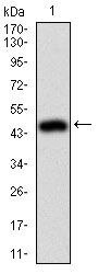
- Western blot analysis of MSN using a MSN monoclonal antibody (Product # MA5-17130) against a human MSN (AA: 292-491) recombinant protein.
- Submitted by
- Invitrogen Antibodies (provider)
- Main image
- Experimental details
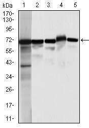
- Western blot analysis of MSN using MSN monoclonal antibody (Product # MA5-17130) in HeLa (1), A431 (2), Jurkat (3), HEK293 (4), and COS-7 (5) cell lysate.

- Submitted by
- Invitrogen Antibodies (provider)
- Main image
- Experimental details
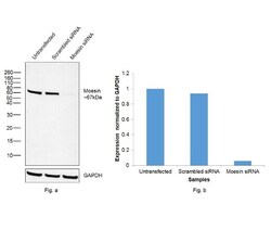
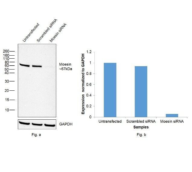
- Knockdown of Moesin was achieved by transfecting A549 cells with Moesin specific siRNAs (Silencer® select Product # s8986). Western blot analysis (Fig. a) was performed using whole cell extracts from Moesin knockdown cells (lane 3), non-specific scrambled siRNA transfected cells (lane 2) and untransfected cells (lane 1). The blots were probed using Moesin Monoclonal Antibody (2C12) (Product # MA5-17130, 1:1000 dilution) and Goat Anti-Mouse IgG Secondary Antibody, HRP conjugate (Product # A28177, 1:4000 dilution). Densitometric analysis of this western blot is shown in histogram (Fig. b). Decrease in signal upon siRNA mediated knock down confirms that antibody is specific to Moesin.
- Submitted by
- Invitrogen Antibodies (provider)
- Main image
- Experimental details
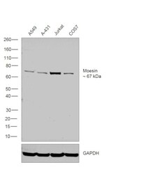
- Western blot was performed using Moesin Monoclonal Antibody (2C12) (Product # MA5-17130) and a 67 kDa band corresponding to Moesin was observed across cell lines tested. Whole cell extracts (30 µg lysate) of A549 (Lane 1), A-431 (Lane 2), Jurkat (Lane 3) and COS7 (Lane 4) were electrophoresed using Novex® NuPAGE® 4-12 % Bis-Tris gel (Product # NP0321BOX). Resolved proteins were then transferred onto a nitrocellulose membrane (Product # IB23001) by iBlot® 2 Dry Blotting System (Product # IB21001). The blot was probed with the primary antibody (1:1000 dilution) and detected by chemiluminescence using Goat Anti-Mouse IgG Secondary Antibody, HRP conjugate (Product # A28177, 1:4000 dilution) using the iBright FL 1000 (Product # A32752). Chemiluminescent detection was performed using Novex® ECL Chemiluminescent Substrate Reagent Kit (Product # WP20005).

- Submitted by
- Invitrogen Antibodies (provider)
- Main image

- Experimental details
- Western blot analysis of MSN using a MSN monoclonal antibody (Product # MA5-17130) against a human MSN (AA: 292-491) recombinant protein.

- Submitted by
- Invitrogen Antibodies (provider)
- Main image

- Experimental details
- Western blot analysis of MSN using MSN monoclonal antibody (Product # MA5-17130) in HeLa (1), A431 (2), Jurkat (3), HEK293 (4), and COS-7 (5) cell lysate.

- Submitted by
- Invitrogen Antibodies (provider)
- Main image
- Experimental details
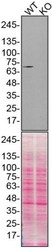
- Western blot analysis of Moesin was performed by loading 25 µg of WT (lane 1) and MSN CRISPR KO (lane 2) HeLa cell lysates in RIPA buffer onto a 4-15% gradient polyacrylamide gel. Proteins were transferred to nitrocellulose membrane and blocked in 5% milk. Ponceau stained transfer of blot is shown. Moesin was detected at approximately 68 kDa using a Moesin monoclonal antibody (Product # MA5-17130) at a dilution of 1:1,000 in 5% BSA in TBST overnight at 4 deg, followed by secondary antibody diluted to 0.2 µg/mL using Goat anti-Mouse IgG (H+L) HRP antibody (Product # 62-6520). Chemiluminescent detection was performed using Pierce ECL Western Blotting Substrate (Product # 32106). Data courtesy of YCharOS Inc., an open science company with the mission of characterizing commercially available antibodies using knockout validation.

Supportive validation
- Submitted by
- Invitrogen Antibodies (provider)
- Main image
- Experimental details
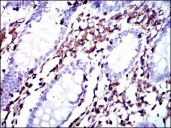
- Immunohistochemical analysis of paraffin-embedded colon tissues using MSN monoclonal antibody (Product # MA5-17130) followed with DAB staining.

Supportive validation
- Submitted by
- Invitrogen Antibodies (provider)
- Main image
- Experimental details
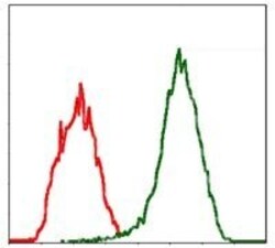
- Flow cytometric analysis of Jurkat cells using MSN monoclonal antibody (Product # MA5-17130) (green) and negative control (red).

Supportive validation
- Submitted by
- Invitrogen Antibodies (provider)
- Main image

- Experimental details
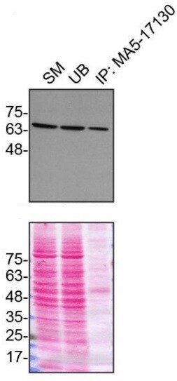
- Immunoprecipitation of Moesin was performed on HeLa cell lysates. Antibody-bead conjugates were prepared by adding 1 µg of Moesin monoclonal antibody (Product # MA5-17130) with 30 µL of protein G-Sepharose beads and rocked overnight at 4°C. 1 µg of MSN KO lysate was incubated with antibody-bead conjugate for 2 hrs at 4°C. After multiple washes, 10% starting material (SM), 10% unbound fraction (UB) and immunoprecipitated fraction (IP) were processed for immunoblot using a Moesin monoclonal antibody. Ponceau stained transfer of blot is shown. Data courtesy of YCharOS Inc., an open science company with the mission of characterizing commercially available antibodies using knockout validation.