 Explore
 Validate
 Learn
Learn Western blot
 Immunocytochemistry
Antibody data
- Antibody Data
- Antigen structure
- References [0]
- Comments [0]
- Validations
- Western blot [3]
- Immunohistochemistry [4]
- Flow cytometry [1]
Submit
Validation data
Reference
Comment
Report error
- Product number
- NBP2-32875 - Provider product page

- Provider
- Novus Biologicals
- Product name
- Mouse Monoclonal Moesin Antibody
- Antibody type
- Monoclonal
- Description
- Protein A purified. Recognizes 78kDa moesin protein. Moesin, a member of the talin-4.1 superfamily, is a linking protein of the sub-membranous actin cytoskeleton. It is expressed in variable amounts in cells of different phenotypes such as macrophages, lymphocytes, fibroblastic, endothelial, epithelial, and neuronal cell lines but not in blood cells. The ERM proteins, ezrin, radixin, and moesin are involved in a variety of cellular functions, such as cell adhesion, migration, and the organization of cell surface structures, and are highly homologous, both in protein sequence and in functional activity, with merlin/schwannomin, a neurofibromatosis-2-associated tumor-suppressor protein. Cell lines of epithelial and mesothelial origin contain both moesin and radixin whereas cells of endothelial and lymphoid origin express moesin.
- Reactivity
- Human, Rat
- Host
- Mouse
- Isotype
- IgG
- Vial size
- 100 ug
- Concentration
- 0.2 mg/ml
- Storage
- Store at 4C.
No comments: Submit comment
Supportive validation
- Submitted by
- Novus Biologicals (provider)
- Main image

- Experimental details
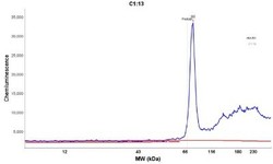
- Simple Western: Moesin Antibody (MSN/491) [NBP2-32875] - Simple Western lane view shows a specific band for Moesin in 0.1 mg/ml of HUVEC lysate. This experiment was performed under reducing conditions using the 12-230 kDa separation system.

- Submitted by
- Novus Biologicals (provider)
- Main image
- Experimental details
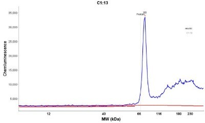
- Simple Western: Moesin Antibody (MSN/491) [NBP2-32875] - Electropherogram image of the corresponding Simple western lane view. Moesin antibody was used at 1 ug/ml dilution on HUVEC lysate(s) respectively.
- Submitted by
- Novus Biologicals (provider)
- Main image
- Experimental details
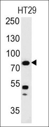
- Western Blot: Moesin Antibody (MSN/491) [NBP2-32875] - Analysis using the Azide and BSA Free version of NBP2-32875. Detection of Moesin in human HT29 Cells.

Supportive validation
- Submitted by
- Novus Biologicals (provider)
- Main image
- Experimental details
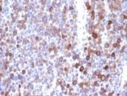
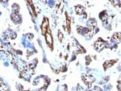
- Immunohistochemistry-Paraffin: Moesin Antibody (MSN/491) [NBP2-32875] - Formalin-paraffin human Melanoma stained with Moesin Mab (MSN/491)

- Submitted by
- Novus Biologicals (provider)
- Main image

- Experimental details
- Immunohistochemistry-Paraffin: Moesin Antibody (MSN/491) [NBP2-32875] - Formalin-paraffin human placenta stained with Moesin MAb (MSN/491)
- Submitted by
- Novus Biologicals (provider)
- Main image

- Experimental details
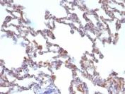
- Immunohistochemistry-Paraffin: Moesin Antibody (MSN/491) [NBP2-32875] - Formalin-paraffin Rat Lung stained with Moesin MAb (MSN/491)
- Submitted by
- Novus Biologicals (provider)
- Main image
- Experimental details
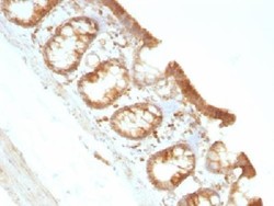
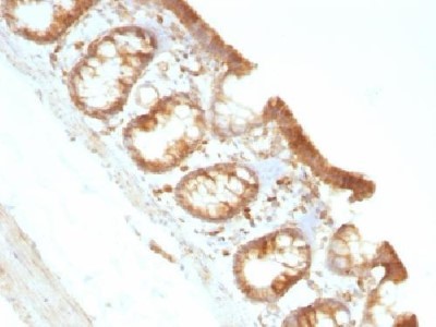
- Immunohistochemistry-Paraffin: Moesin Antibody (MSN/491) [NBP2-32875] - Fomalin-paraffin Rat colon stained with Moesin MAb (MSN/491)
Supportive validation
- Submitted by
- Novus Biologicals (provider)
- Main image
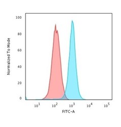
- Experimental details
- Flow Cytometry: Moesin Antibody (MSN/491) [NBP2-32875] - Flow Cytometric Analysis of K562 cells using Moesin Antibody (MSN/491) followed by Goat anti-Mouse IgG-CF488 (Blue); Isotype Control (Red).
