Explore
 Validate
 Learn
Learn Western blot
 Immunocytochemistry
Antibody data
- Antibody Data
- Antigen structure
- References [0]
- Comments [0]
- Validations
- Western blot [5]
- Immunohistochemistry [1]
Submit
Validation data
Reference
Comment
Report error
- Product number
- NBP1-31069 - Provider product page

- Provider
- Novus Biologicals
- Proper citation
- Novus Cat#NBP1-31069, RRID:AB_2272915
- Product name
- Rabbit Polyclonal VPS35 Antibody
- Antibody type
- Polyclonal
- Description
- Antigen Affinity-purified.
- Reactivity
- Human, Mouse, Rat
- Host
- Rabbit
- Isotype
- IgG
- Vial size
- 0.1 ml
- Storage
- Store at 4C short term. Aliquot and store at -20C long term. Avoid freeze-thaw cycles.
No comments: Submit comment
Supportive validation
- Submitted by
- Novus Biologicals (provider)
- Main image
- Experimental details
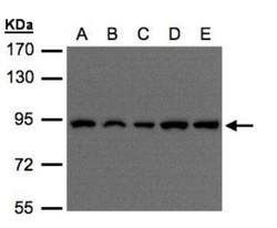
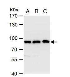
- Western Blot: VPS35 Antibody [NBP1-31069] - Sample(30 ug of whole cell lysate)A:H1299B:HeLa S3 C:Hep G2 D:MOLT4 E:Raji 7. 5% SDS PAGE, antibody diluted at 1:500.
- Submitted by
- Novus Biologicals (provider)
- Main image
- Experimental details
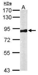
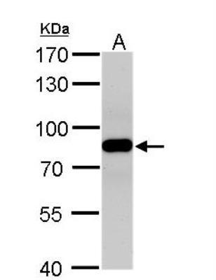
- Western Blot: VPS35 Antibody [NBP1-31069] - Sample (50 ug of whole cell lysate) A: Mouse brain 7. 5% SDS PAGE; antibody diluted at 1:1000.

- Submitted by
- Novus Biologicals (provider)
- Main image
- Experimental details
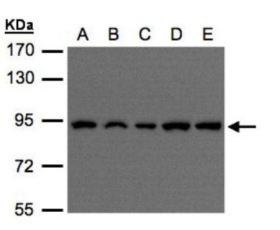
- Western Blot: VPS35 Antibody [NBP1-31069] - A. 30 ug A549 whole cell extract B. 30 ug H1299 whole cell extract C. 30 ug HCT116 whole cell extract 7.5% SDS-PAGE VPS35 antibody [C3], C-term dilution: 1:1000 The HRP-conjugated anti-rabbit IgG antibody (NBP2-19301) was used to detect the primary antibody.

- Submitted by
- Novus Biologicals (provider)
- Main image

- Experimental details
- Western Blot: VPS35 Antibody [NBP1-31069] - A. 50 ug rat brain lysate/extract 7.5% SDS-PAGE VPS35 antibody dilution: 1:500 The HRP-conjugated anti-rabbit IgG antibody (NBP2-19301) was used to detect the primary antibody.
- Submitted by
- Novus Biologicals (provider)
- Main image
- Experimental details
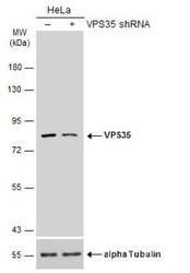
- Western Blot: VPS35 Antibody [NBP1-31069] - Non-transfected (-) and transfected (+) HeLa whole cell extracts (30 ug) were separated by 7.5% SDS-PAGE, and the membrane was blotted with VPS35 antibody diluted at 1:5000. HRP-conjugated anti-rabbit IgG antibody was used to detect the primary antibody.

Supportive validation
- Submitted by
- Novus Biologicals (provider)
- Main image

- Experimental details
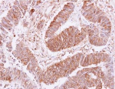
- Immunohistochemistry-Paraffin: VPS35 Antibody [NBP1-31069] - VPS35 antibody [C3], C-term detects VPS35 protein at cytoplasm and membrane on human colon carcinoma by immunohistochemical analysis. Sample: Paraffin-embedded colon carcinoma. VPS35 antibody [C3], C-term dilution: 1:250.