Explore
 Validate
 Learn
Learn Western blot
Antibody data
- Antibody Data
- Antigen structure
- References [7]
- Comments [0]
- Validations
- Western blot [4]
- Immunocytochemistry [2]
- Flow cytometry [1]
- Other assay [3]
Submit
Validation data
Reference
Comment
Report error
- Product number
- MA1-2000 - Provider product page

- Provider
- Invitrogen Antibodies
- Product name
- alpha Catenin Monoclonal Antibody (1G5)
- Antibody type
- Monoclonal
- Antigen
- Purifed from natural sources
- Description
- MA1-2000 detects alpha catenin from human samples.
- Antibody clone number
- 1G5
- Concentration
- 1 mg/mL
Submitted references Exclusion from spheroid formation identifies loss of essential cell-cell adhesion molecules in colon cancer cells.
Leptospira interrogans causes quantitative and morphological disturbances in adherens junctions and other biological groups of proteins in human endothelial cells.
Integration of left-right Pitx2 transcription and Wnt signaling drives asymmetric gut morphogenesis via Daam2.
Characterization of cadherin-24, a novel alternatively spliced type II cadherin.
Mechanism of extracellular domain-deleted dominant negative cadherins.
Expression of N-cadherin by human squamous carcinoma cells induces a scattered fibroblastic phenotype with disrupted cell-cell adhesion.
Identification of plakoglobin domains required for association with N-cadherin and alpha-catenin.
Stadler M, Scherzer M, Walter S, Holzner S, Pudelko K, Riedl A, Unger C, Kramer N, Weil B, Neesen J, Hengstschläger M, Dolznig H
Scientific reports 2018 Jan 18;8(1):1151
Scientific reports 2018 Jan 18;8(1):1151
Leptospira interrogans causes quantitative and morphological disturbances in adherens junctions and other biological groups of proteins in human endothelial cells.
Sato H, Coburn J
PLoS neglected tropical diseases 2017 Jul;11(7):e0005830
PLoS neglected tropical diseases 2017 Jul;11(7):e0005830
Integration of left-right Pitx2 transcription and Wnt signaling drives asymmetric gut morphogenesis via Daam2.
Welsh IC, Thomsen M, Gludish DW, Alfonso-Parra C, Bai Y, Martin JF, Kurpios NA
Developmental cell 2013 Sep 30;26(6):629-44
Developmental cell 2013 Sep 30;26(6):629-44
Characterization of cadherin-24, a novel alternatively spliced type II cadherin.
Katafiasz BJ, Nieman MT, Wheelock MJ, Johnson KR
The Journal of biological chemistry 2003 Jul 25;278(30):27513-9
The Journal of biological chemistry 2003 Jul 25;278(30):27513-9
Mechanism of extracellular domain-deleted dominant negative cadherins.
Nieman MT, Kim JB, Johnson KR, Wheelock MJ
Journal of cell science 1999 May;112 ( Pt 10):1621-32
Journal of cell science 1999 May;112 ( Pt 10):1621-32
Expression of N-cadherin by human squamous carcinoma cells induces a scattered fibroblastic phenotype with disrupted cell-cell adhesion.
Islam S, Carey TE, Wolf GT, Wheelock MJ, Johnson KR
The Journal of cell biology 1996 Dec;135(6 Pt 1):1643-54
The Journal of cell biology 1996 Dec;135(6 Pt 1):1643-54
Identification of plakoglobin domains required for association with N-cadherin and alpha-catenin.
Sacco PA, McGranahan TM, Wheelock MJ, Johnson KR
The Journal of biological chemistry 1995 Aug 25;270(34):20201-6
The Journal of biological chemistry 1995 Aug 25;270(34):20201-6
No comments: Submit comment
Supportive validation
- Submitted by
- Invitrogen Antibodies (provider)
- Main image

- Experimental details
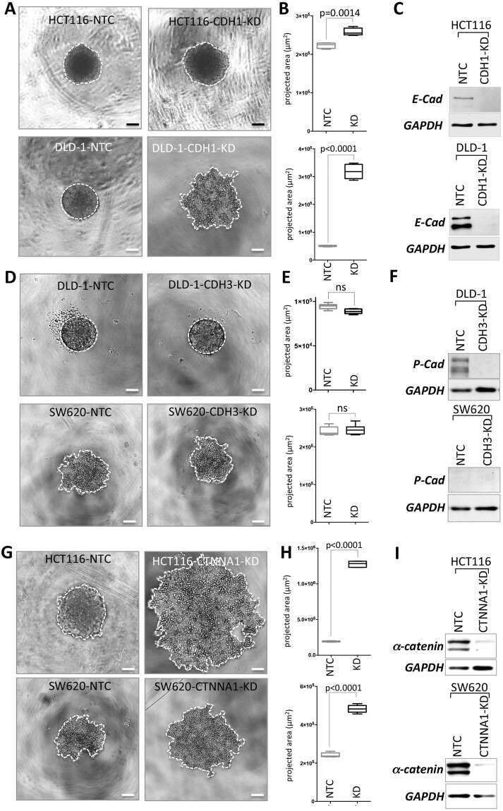
- Western blot analysis of alpha Catenin was performed by loading 25 µg of Hela (lane 1), A431 (lane 2) and mouse brain (lane 3) onto an SDS polyacrylamide gel. Proteins were transferred to a PVDF membrane and blocked at 4ºC overnight. The membrane was probed with an Alpha Catenin monoclonal antibody (Product # MA1-2000) at a dilution of 1:500 overnight at 4°C, washed in TBST, and probed with an HRP-conjugated secondary antibody for 1 hr at room temperature in the dark. Chemiluminescent detection was performed using Pierce ECL Plus Western Blotting Substrate (Product # 32132). Results show a band at ~102 kDa in human cell lines, and no reactivity with mouse.
- Submitted by
- Invitrogen Antibodies (provider)
- Main image
- Experimental details
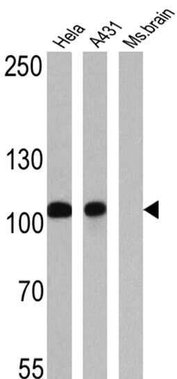
- Western blot analysis was performed on whole cell extracts (30 µg lysate) of HeLa (Lane 1), A-431 (Lane 2) and MCF7 (Lane 3). The blots were probed with Anti-alpha Catenin Mouse Monoclonal Antibody (Product # MA1-2000, 1:250-1:750 dilution) and detected by chemiluminescence using Goat anti-Mouse IgG (H+L) Secondary Antibody, HRP conjugate (Product # 62-6520, 1:4000 dilution). A 100 kDa bands corresponding to alpha Catenin was observed across cell lines tested. Known quantity of protein samples were electrophoresed using Novex® NuPAGE® 4-12 % Bis-Tris gel (Product # NP0322BOX), XCell SureLock™ Electrophoresis System (Product # EI0002) and Novex® Sharp Pre-Stained Protein Standard (Product # LC5800). Resolved proteins were then transferred onto a nitrocellulose membrane with iBlot® 2 Dry Blotting System (Product # IB21001). The membrane was probed with the relevant primary and secondary Antibody following blocking with 5 % skimmed milk. Chemiluminescent detection was performed using Pierce™ ECL Western Blotting Substrate (Product # 32106).

- Submitted by
- Invitrogen Antibodies (provider)
- Main image
- Experimental details
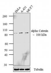
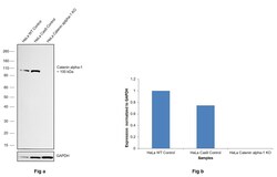
- Knockdown of alpha Catenin was achieved by transfecting A-431 with alpha Catenin specific siRNAs (Silencer® select Product # s3717, s3716). Western blot analysis (Fig. a) was performed using membrane extracts from the alpha Catenin knockdown cells (lane 3), non-specific scrambled siRNA transfected cells (lane 2) and untransfected cells (lane 1). The blots were probed with alpha Catenin Monoclonal Antibody (1G5) (Product # MA1-2000, 1:1000 dilution) and Goat anti-Mouse IgG (H+L) Superclonal™ Secondary Antibody, HRP conjugate (Product # A28177, 0.25 µg/mL, 1:4000 dilution). Densitometric analysis of this western blot is shown in histogram (Fig. b). Absence of signal upon siRNA mediated knock down confirms that antibody is specific to alpha Catenin.

- Submitted by
- Invitrogen Antibodies (provider)
- Main image
- Experimental details
- Knockout of Catenin alpha-1 was achieved by CRISPR-Cas9 genome editing using LentiArray™ Lentiviral sgRNA (Product # A32042, Assay ID CRISPR787281_LV) and LentiArray Cas9 Lentivirus (Product # A32064). Western blot analysis of Catenin alpha-1 was performed by loading 30 µg of HeLa Wild Type (Lane 1), HeLa Cas9 (Lane 2) andHeLa Catenin alpha-1 KO (Lane 3) whole cell extracts. The samples were electrophoresed using NuPAGE™ Novex™ 4-12% Bis-Tris Protein Gel (Product # NP0322BOX). Resolved proteins were then transferred onto a nitrocellulose membrane (Product # IB23001) by iBlot® 2 Dry Blotting System (Product # IB21001). The blot was probed with Anti-alpha Catenin Monoclonal Antibody (1G5) (Product # MA1-2000, 1:1,000 dilution) and Goat anti-Mouse IgG (H+L) Superclonal™ Recombinant Secondary Antibody, HRP (Product # A28177, 1:5,000 dilution) using the iBright FL 1000 (Product # A32752). Chemiluminescent detection was performed using Novex® ECL Chemiluminescent Substrate Reagent Kit (Product # WP20005). Loss of signal upon CRISPR mediated knockout (KO) using the LentiArray™ CRISPR product line confirms that antibody is specific to Catenin alpha-1.

Supportive validation
- Submitted by
- Invitrogen Antibodies (provider)
- Main image
- Experimental details
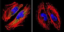
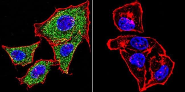
- Immunofluorescent analysis of alpha Catenin using Catenin alpha Monoclonal Antibody (1G5) (Product # MA1-2000) shows staining in A2058 Cells. Catenin alpha (green), F-Actin staining with Phalloidin (red) and nuclei with DAPI (blue) is shown. Cells were grown on chamber slides and fixed with formaldehyde prior to staining. Cells were probed without (control) or with an antibody recognizing Catenin alpha (Product # MA1-2000) at a dilution of 1:20 over night at 4 °C, washed with PBS and incubated with a DyLight-488 conjugated secondary antibody (Product # 35552 for GAR, Product # 35503 for GAM). Images were taken at 60X magnification.
- Submitted by
- Invitrogen Antibodies (provider)
- Main image

- Experimental details
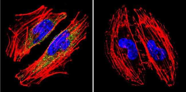
- Immunofluorescent analysis of alpha Catenin using Catenin alpha Monoclonal Antibody (1G5) (Product # MA1-2000) shows staining in Hela Cells. Catenin alpha (green), F-Actin staining with Phalloidin (red) and nuclei with DAPI (blue) is shown. Cells were grown on chamber slides and fixed with formaldehyde prior to staining. Cells were probed without (control) or with an antibody recognizing Catenin alpha (Product # MA1-2000) at a dilution of 1:20 over night at 4 °C, washed with PBS and incubated with a DyLight-488 conjugated secondary antibody (Product # 35552 for GAR, Product # 35503 for GAM). Images were taken at 60X magnification.
Supportive validation
- Submitted by
- Invitrogen Antibodies (provider)
- Main image
- Experimental details
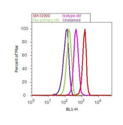
- Flow cytometry analysis of alpha Catenin was done on A431 cells. Cells were fixed with 70% ethanol for 10 minutes, permeabilized with 0.25% Triton™ X-100 for 20 minutes, and blocked with 5% BSA for 30 minutes at room temperature. Cells were labeled with alpha Catenin Mouse Monoclonal Antibody (MA12000, red histogram) or with mouse isotype control (pink histogram) at 3-5 ug/million cells in 2.5% BSA. After incubation at room temperature for 2 hours, the cells were labeled with Alexa Fluor® 488 Rabbit Anti-Mouse Secondary Antibody (A11059) at a dilution of 1:400 for 30 minutes at room temperature. The representative 10,000 cells were acquired and analyzed for each sample using an Attune® Acoustic Focusing Cytometer. The purple histogram represents unstained control cells and the green histogram represents no-primary-antibody control.

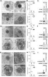
Supportive validation
- Submitted by
- Invitrogen Antibodies (provider)
- Main image
- Experimental details
- NULL
- Submitted by
- Invitrogen Antibodies (provider)
- Main image

- Experimental details
- NULL
- Submitted by
- Invitrogen Antibodies (provider)
- Main image
- Experimental details
- NULL