 Explore
 Validate
 Learn
Learn Western blot
 Immunohistochemistry
Antibody data
- Antibody Data
- Antigen structure
- References [0]
- Comments [0]
- Validations
- Immunohistochemistry [2]
Submit
Validation data
Reference
Comment
Report error
- Product number
- BAF233 - Provider product page

- Provider
- R&D Systems
- Product name
- Human FGF basic/FGF2/bFGF Biotinylated Antibody
- Antibody type
- Polyclonal
- Description
- Antigen Affinity-purified. Detects human FGF basic/FGF2/bFGF in Western blots. In Western blots, approximately 10% cross-reactivity with recombinant human (rh) FGF acidic and rhFGF-7 is observed under non-reducing conditions.
- Reactivity
- Human
- Host
- Goat
- Conjugate
- Biotin
- Antigen sequence
P09038- Isotype
- IgG
- Vial size
- 50 ug
- Concentration
- LYOPH
- Storage
- Use a manual defrost freezer and avoid repeated freeze-thaw cycles. 12 months from date of receipt, -20 to -70 °C as supplied. 1 month, 2 to 8 °C under sterile conditions after reconstitution. 6 months, -20 to -70 °C under sterile conditions after reconstitution.
No comments: Submit comment
Supportive validation
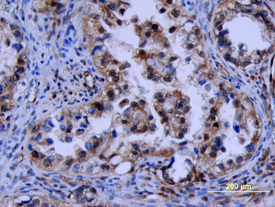
- Submitted by
- R&D Systems (provider)
- Main image

- Experimental details
- FGF basic/FGF2 in Human Ovarian Cancer Tissue. FGF basic/FGF2 was detected in immersion fixed paraffin-embedded sections of human ovarian cancer tissue using Human FGF basic/FGF2 Biotinylated Antigen Affinity-purified Polyclonal Antibody (Catalog # BAF233) at 10 µg/mL overnight at 4 °C. Tissue was stained using the Anti-Goat HRP-DAB Cell & Tissue Staining Kit (brown; Catalog # CTS008) and counterstained with hematoxylin (blue). View our protocol for Chromogenic IHC Staining of Paraffin-embedded Tissue Sections.
- Submitted by
- R&D Systems (provider)
- Main image
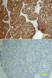
- Experimental details
- FGF basic/FGF2 in Human Ovary. FGF basic/FGF2 was detected in immersion fixed paraffin-embedded sections of human ovarian array using Human FGF basic/FGF2 Biotinylated Antigen Affinity-purified Polyclonal Antibody (Catalog # BAF233) at 10 µg/mL overnight at 4 °C. Tissue was stained using the Anti-Goat HRP-DAB Cell & Tissue Staining Kit (brown; Catalog # CTS008) and counterstained with hematoxylin (blue). Lower panel shows a lack of labeling if primary antibodies are omitted and tissue is stained only with secondary antibody followed by incubation with detection reagents. View our protocol for Chromogenic IHC Staining of Paraffin-embedded Tissue Sections.
