Explore
 Validate
 Learn
Learn Western blot
 Immunohistochemistry
Antibody data
- Antibody Data
- Antigen structure
- References [1]
- Comments [0]
- Validations
- Western blot [1]
- Immunocytochemistry [1]
- Other assay [1]
Submit
Validation data
Reference
Comment
Report error
- Product number
- OSB00018G - Provider product page

- Provider
- Invitrogen Antibodies
- Product name
- BDNF Polyclonal Antibody
- Antibody type
- Polyclonal
- Antigen
- Synthetic peptide
- Description
- Fixation: Samples can be stored in IC Fixation Buffer (cat. 00-8222) (100 µL of cell sample + 100 µL of IC Fixation Buffer) or 1-step Fix/Lyse Solution (cat. 00-5333) for up to 3 days in the dark at 4°C with minimal impact on brightness and FRET efficiency/compensation. Some generalizations regarding fluorophore performance after fixation can be made, but clone specific performance should be determined empirically.
- Reactivity
- Human, Mouse, Rat
- Host
- Rabbit
- Isotype
- IgG
- Vial size
- 500 µg
- Concentration
- 1 mg/mL
- Storage
- Store at 4°C short term. For long term storage, store at -20°C, avoiding freeze/thaw cycles. Glycerol (1:1) may be added for added stability.
Submitted references Modafinil Reduces Neuronal Pyroptosis and Cognitive Decline After Sleep Deprivation.
Xiong X, Zuo Y, Cheng L, Yin Z, Hu T, Guo M, Han Z, Ge X, Li W, Wang Y, Wang D, Wang C, Zhang L, Zhang Y, Liu Q, Chen F, Lei P
Frontiers in neuroscience 2022;16:816752
Frontiers in neuroscience 2022;16:816752
No comments: Submit comment
Supportive validation
- Submitted by
- Invitrogen Antibodies (provider)
- Main image
- Experimental details
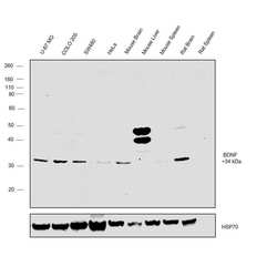
- Western blot was performed using Anti-BDNF Polyclonal Antibody (Product # OSB00018G) and a 34kDa band corresponding to Brain-derived neurotrophic factor was observed across the panel tested except for Mouse liver and spleen and Rat Spleen which are reported to be negative. Whole cell extracts and tissue extracts (30 µg lysate) of U-87 MG (Lane 1), COLO 205 (Lane 2), SW480 (Lane 3), HeLa (Lane 4), Mouse Brain (Lane 5), Mouse Liver (Lane 6), Mouse Spleen (Lane 7), Rat Brain (Lane 8), Rat Spleen (Lane 9) were electrophoresed using NuPAGE™ 4-12% Bis-Tris Protein Gel (Product # NP0322BOX). Resolved proteins were then transferred onto a Nitrocellulose membrane (Product # IB23001) by iBlot® 2 Dry Blotting System (Product # IB21001). The blot was probed with the primary antibody (1:500 dilution) and detected by chemiluminescence with Goat anti-Rabbit IgG (H+L) Superclonal™ Recombinant Secondary Antibody, HRP (Product # A27036, 1:4000 dilution) using the iBright FL 1000 (Product # A32752). Chemiluminescent detection was performed using Novex® ECL Reagent Kit (Product # WP20005).
Supportive validation
- Submitted by
- Invitrogen Antibodies (provider)
- Main image
- Experimental details
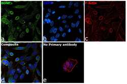
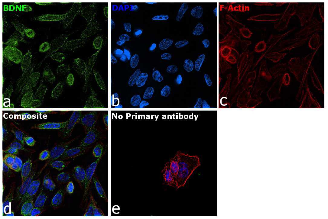
- Immunofluorescence analysis of Brain-derived neurotrophic factor was performed using 70% confluent log phase SW480 cells. The cells were fixed with 4% paraformaldehyde for 10 minutes, permeabilized with 0.1% Triton™ X-100 for 15 minutes, and blocked with 2% BSA for 45 minutes at room temperature. The cells were labeled with BDNF Polyclonal Antibody (Product # OSB00018G) at 1:200 dilution in 0.1% BSA, incubated at 4 degree celsius overnight and then labeled with Goat anti-Rabbit IgG (H+L) Highly Cross-Adsorbed Secondary Antibody, Alexa Fluor Plus 488 (Product # A32731), (1:3000 dilution), for 45 minutes at room temperature (Panel a: Green). Nuclei (Panel b:Blue) were stained with ProLong™ Diamond Antifade Mountant with DAPI (Product # P36962). F-actin (Panel c: Red) was stained with Rhodamine Phalloidin (Product # R415, 1:300). Panel d represents the merged image showing cytoplasmic localization. Panel e represents control cells with no primary antibody to assess background. The images were captured at 60X magnification.
Supportive validation
- Submitted by
- Invitrogen Antibodies (provider)
- Main image
- Experimental details
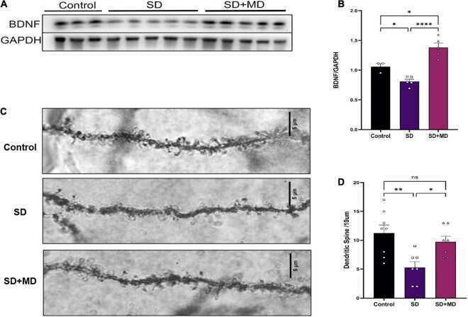
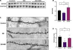
- FIGURE 6 Modafinil decreased BDNF expression and alleviated dendritic spine loss in hippocampal CA3 pyramidal neurons in mice subjected to SD. (A,B) Western bot analysis of BDNF expression in the hippocampus. All data presented are means +- SD; N = 3-5 mice per group. (C,D) The density of CA3 pyramidal neurons, as measured by Golgi staining, was decreased in mice subjected to SD, and modafinil reversed this decrease. All data presented are means +- SD; N = 7-8 mice per group. * P < 0.05, ** P < 0.01, **** P < 0.0001.