Explore
 Validate
 Learn
Learn Western blot
Antibody data
- Antibody Data
- Antigen structure
- References [4]
- Comments [0]
- Validations
- Western blot [1]
- Other assay [3]
Submit
Validation data
Reference
Comment
Report error
- Product number
- 44-612G - Provider product page

- Provider
- Invitrogen Antibodies
- Product name
- Phospho-ERK5 (Thr218, Tyr220) Polyclonal Antibody
- Antibody type
- Polyclonal
- Antigen
- Synthetic peptide
- Reactivity
- Human
- Host
- Rabbit
- Isotype
- IgG
- Vial size
- 100 µL
- Storage
- -20°C
Submitted references Dexmedetomidine and Netrin-1 Combination Therapy Inhibits Endoplasmic Reticulum Stress by Regulating the ERK5/MEF2A Pathway to Attenuate Cerebral Ischemia Injury.
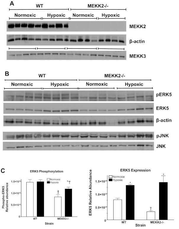
MAP kinase kinase kinase-2 (MEKK2) regulates hypertrophic remodeling of the right ventricle in hypoxia-induced pulmonary hypertension.
Abl-kinase-sensitive levels of ERK5 and its intrinsic basal activity contribute to leukaemia cell survival.
Phosphotyrosine-specific phosphatase PTP-SL regulates the ERK5 signaling pathway.
Yin JW, Li J, Ren YM, Li Y, Wang RX, Wang S, Zuo YX
Frontiers in neuroscience 2021;15:641345
Frontiers in neuroscience 2021;15:641345
MAP kinase kinase kinase-2 (MEKK2) regulates hypertrophic remodeling of the right ventricle in hypoxia-induced pulmonary hypertension.
Brown RD, Ambler SK, Li M, Sullivan TM, Henry LN, Crossno JT Jr, Long CS, Garrington TP, Stenmark KR
American journal of physiology. Heart and circulatory physiology 2013 Jan 15;304(2):H269-81
American journal of physiology. Heart and circulatory physiology 2013 Jan 15;304(2):H269-81
Abl-kinase-sensitive levels of ERK5 and its intrinsic basal activity contribute to leukaemia cell survival.
Buschbeck M, Hofbauer S, Di Croce L, Keri G, Ullrich A
EMBO reports 2005 Jan;6(1):63-9
EMBO reports 2005 Jan;6(1):63-9
Phosphotyrosine-specific phosphatase PTP-SL regulates the ERK5 signaling pathway.
Buschbeck M, Eickhoff J, Sommer MN, Ullrich A
The Journal of biological chemistry 2002 Aug 16;277(33):29503-9
The Journal of biological chemistry 2002 Aug 16;277(33):29503-9
No comments: Submit comment
Supportive validation
- Submitted by
- Invitrogen Antibodies (provider)
- Main image
- Experimental details
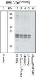
- Extracts prepared from HEK293 cells transiently transfected with plasmids expressing ERK5 kinase domain (ERK5kin) and constitutively activated MEK5D-D were resolved by SDS-PAGE on a 10% polyacrylamide gel and transferred to PVDF. Membranes were blocked with a 5% BSA-TBST buffer overnight at 4°C, then were incubated with the ERK5 (Thr218, Tyr220) antibody for two hours at room temperature in a 3% BSA-TBST buffer, following prior incubation with: no peptide (1), the non-phosphopeptide corresponding to the immunogen (2), a generic phosphothreonine-containing peptide (3), a generic phosphotyrosine-containing peptide (4), the phosphopeptide derived from the corresponding region of ERK1&2 (5), or, the phosphopeptide immunogen (6). After washing, membranes were incubated with goat F (ab’)2 anti-rabbit IgG alkaline phosphatase conjugate (Product # ALI4405) and bands were detected using the Tropix WesternStar™ detection method. The data show that while there is some cross-reactivity with ERK1&2, only the phosphopeptide corresponding to ERK5 (pTpY218/220) completely blocks the antibody signal, demonstrating the specificity of the antibody. NOTE: The antibody signal appears at ~50 kDa as this is the molecular weight of the transiently transfected ERK5 kinase domain.

Supportive validation
- Submitted by
- Invitrogen Antibodies (provider)
- Main image

- Experimental details
- NULL
- Submitted by
- Invitrogen Antibodies (provider)
- Main image

- Experimental details
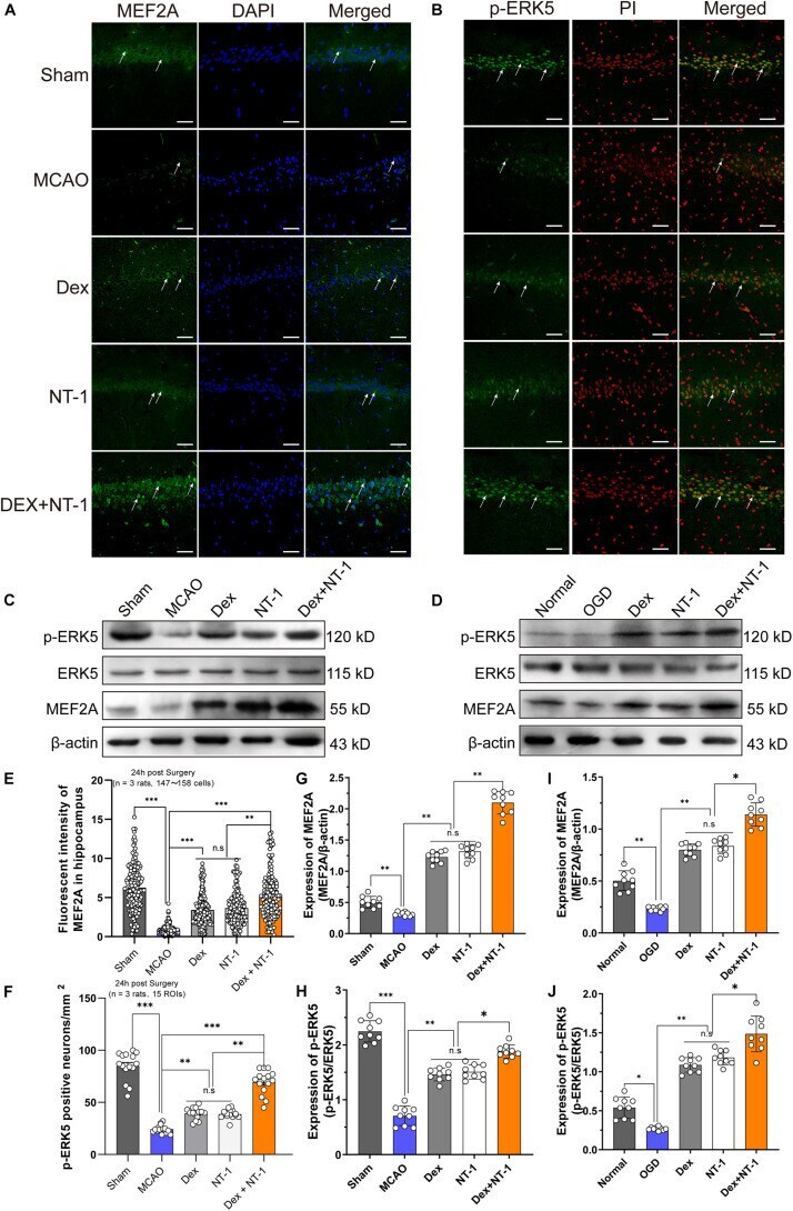
- FIGURE 6 Dexmedetomidine and Netrin-1 up-regulated the expression of the ERK5 and MEF2A after MCAO injury and OGD injury. (A) MEF2A expressions in hippocampus after MCAO injury. Immunofluorescence staining exhibited positive cells of MEF2A. Green indicated MEF2A expression in plasm of pyramidal neurons, and blue indicated the nucleus. Scale bar = 100 mum. (B) p-ERK5 expressions in hippocampus after MCAO injury. Immunofluorescence staining exhibited positive cells of p-ERK5. Green indicated p-ERK5 expression in nucleus of pyramidal neurons, and red indicated the nucleus. Scale bar = 100 mum. (C,D) Western blot analysis of MEF2A, ERK5 and p-ERK5 proteins, beta-actin was used as an internal control. (E-I) Quantitative analysis of MEF2A and GRP78 expression levels. (F-J) Quantitative analysis of p-ERK5 expression levels. * P < 0.05, ** P < 0.01 and *** P < 0.001 using one-way ANOVA followed by Tukey's post hoc tests.
- Submitted by
- Invitrogen Antibodies (provider)
- Main image

- Experimental details
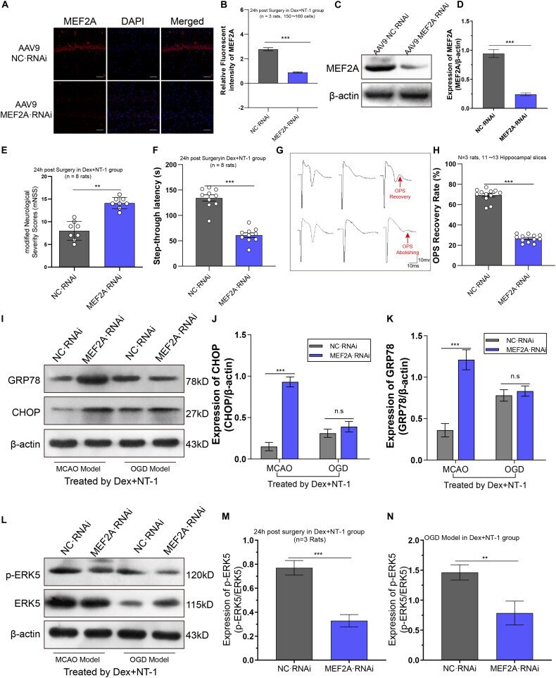
- FIGURE 7 Knockdown MEF2A weakened the therapeutic effect of dexmedetomidine combined with Netrin-1 after MCAO and OGD injuries. (A,B) Immunofluorescence staining exhibited downregulation in MEF2A expression in the hippocampus at 14 days after AAV9-MEF2A RNAi microinjection. (C,D) Western blot analysis shows downregulation in MEF2A expression in the hippocampus at 14 days after AAV9-MEF2A RNAi microinjection. (E) The mNSS result after MEF2A knockdown. (F) The step-through latency result after MEF2A knockdown. (G,H) The OPS recovery rate after MEF2A knockdown. (I-K) Western blot analysis of ERS-related proteins CHOP and GRP78 in the hippocampus at 14 days after AAV9-MEF2A RNAi microinjection, beta-actin was used as an internal control. (L-N) Western blot analysis of ERS-related proteins ERK5 and p-ERK5 in the hippocampus at 14 days after AAV9-MEF2A RNAi microinjection, beta-actin was used as an internal control. ** P < 0.01 and *** P < 0.001 using one-way ANOVA followed by Tukey's post hoc tests.