Explore
 Validate
 Learn
Learn Western blot
Antibody data
- Antibody Data
- Antigen structure
- References [5]
- Comments [0]
- Validations
- Western blot [7]
- Immunocytochemistry [1]
- Immunohistochemistry [2]
Submit
Validation data
Reference
Comment
Report error
- Product number
- GTX112734 - Provider product page

- Provider
- GeneTex
- Proper citation
- GeneTex Cat#GTX112734, RRID:AB_10729183
- Product name
- N-Cadherin antibody [N1N3]
- Antibody type
- Polyclonal
- Reactivity
- Human, Mouse, Rat
- Host
- Rabbit
Submitted references Elevation of YAP promotes the epithelial-mesenchymal transition and tumor aggressiveness in colorectal cancer.
Overexpression of lipocalin 2 in human cervical cancer enhances tumor invasion.
N-cadherin coordinates AMP kinase-mediated lung vascular repair.
Involvement of RARRES3 in the regulation of Wnt proteins acylation and signaling activities in human breast cancer cells.
CD63 tetraspanin is a negative driver of epithelial-to-mesenchymal transition in human melanoma cells.
Ling HH, Kuo CC, Lin BX, Huang YH, Lin CW
Experimental cell research 2017 Jan 1;350(1):218-225
Experimental cell research 2017 Jan 1;350(1):218-225
Overexpression of lipocalin 2 in human cervical cancer enhances tumor invasion.
Chung IH, Wu TI, Liao CJ, Hu JY, Lin YH, Tai PJ, Lai CH, Lin KH
Oncotarget 2016 Mar 8;7(10):11113-26
Oncotarget 2016 Mar 8;7(10):11113-26
N-cadherin coordinates AMP kinase-mediated lung vascular repair.
Jian MY, Liu Y, Li Q, Wolkowicz P, Alexeyev M, Zmijewski J, Creighton J
American journal of physiology. Lung cellular and molecular physiology 2016 Jan 1;310(1):L71-85
American journal of physiology. Lung cellular and molecular physiology 2016 Jan 1;310(1):L71-85
Involvement of RARRES3 in the regulation of Wnt proteins acylation and signaling activities in human breast cancer cells.
Hsu TH, Jiang SY, Chang WL, Eckert RL, Scharadin TM, Chang TC
Cell death and differentiation 2015 May;22(5):801-14
Cell death and differentiation 2015 May;22(5):801-14
CD63 tetraspanin is a negative driver of epithelial-to-mesenchymal transition in human melanoma cells.
Lupia A, Peppicelli S, Witort E, Bianchini F, Carloni V, Pimpinelli N, Urso C, Borgognoni L, Capaccioli S, Calorini L, Lulli M
The Journal of investigative dermatology 2014 Dec;134(12):2947-2956
The Journal of investigative dermatology 2014 Dec;134(12):2947-2956
No comments: Submit comment
Supportive validation
- Submitted by
- GeneTex (provider)
- Main image

- Experimental details
- Sample (30 ?g of whole cell lysate) A: K562 7.5% SDS PAGE GTX112734 diluted at 1:1000 The HRP-conjugated anti-rabbit IgG antibody (GTX213110-01) was used to detect the primary antibody.
- Submitted by
- GeneTex (provider)
- Main image

- Experimental details
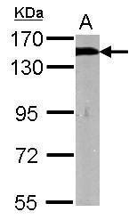
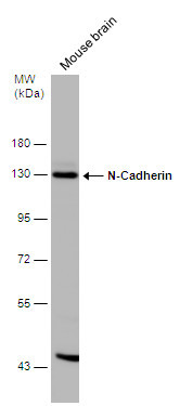
- Sample (50 ug of whole cell lysate) A: Mouse brain 7.5% SDS PAGE GTX112734 diluted at 1:1000
- Validation comment
- WB
- Submitted by
- GeneTex (provider)
- Main image
- Experimental details
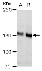
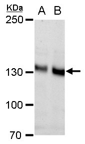
- N-Cadherin antibody [N1N3] detects N-Cadherin protein by western blot analysis.A. 30 ?g PC-12 whole cell extract B. 30 ?g Rat2 whole cell extract5% SDS-PAGEN-Cadherin antibody [N1N3] (GTX112734) dilution: 1:1000 The HRP-conjugated anti-rabbit IgG antibody (GTX213110-01) was used to detect the primary antibody.
- Submitted by
- GeneTex (provider)
- Main image
- Experimental details
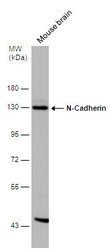
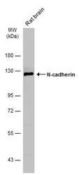
- Mouse tissue extract (50 ?g) was separated by 7.5% SDS-PAGE, and the membrane was blotted with N-Cadherin antibody [N1N3] (GTX112734) diluted at 1:500. The HRP-conjugated anti-rabbit IgG antibody (GTX213110-01) was used to detect the primary antibody.
- Submitted by
- GeneTex (provider)
- Main image
- Experimental details
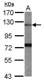
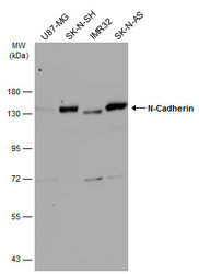
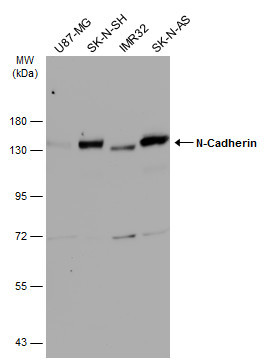
- Various whole cell extracts (30 ?g) were separated by 7.5% SDS-PAGE, and the membrane was blotted with N-Cadherin antibody [N1N3] (GTX112734) diluted at 1:1000. The HRP-conjugated anti-rabbit IgG antibody (GTX213110-01) was used to detect the primary antibody.
- Submitted by
- GeneTex (provider)
- Main image
- Experimental details
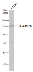
- Whole cell extract (30 ?g) was separated by 7.5% SDS-PAGE, and the membrane was blotted with N-Cadherin antibody [N1N3] (GTX112734) diluted at 1:1000. The HRP-conjugated anti-rabbit IgG antibody (GTX213110-01) was used to detect the primary antibody.

- Submitted by
- GeneTex (provider)
- Main image
- Experimental details
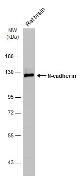
- Rat tissue extract (50 ?g) was separated by 7.5% SDS-PAGE, and the membrane was blotted with N-cadherin antibody (GTX112734) diluted at 1:1000. The HRP-conjugated anti-rabbit IgG antibody (GTX213110-01) was used to detect the primary antibody.
Supportive validation
- Submitted by
- GeneTex (provider)
- Main image
- Experimental details
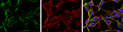
- N-Cadherin antibody [N1N3] detects N-Cadherin protein at cell membrane and cytoplasm by immunofluorescent analysis.Sample: U-87 MG cells were fixed in 4% paraformaldehyde at RT for 15 min.Green: N-Cadherin protein stained by N-Cadherin antibody [N1N3] (GTX112734) diluted at 1:250.Red: beta Tubulin 3/ TUJ1 protein stained by beta Tubulin 3/ TUJ1 antibody (GTX631836) diluted at 1:200.Blue: Hoechst 33342 staining.

Supportive validation
- Submitted by
- GeneTex (provider)
- Main image
- Experimental details
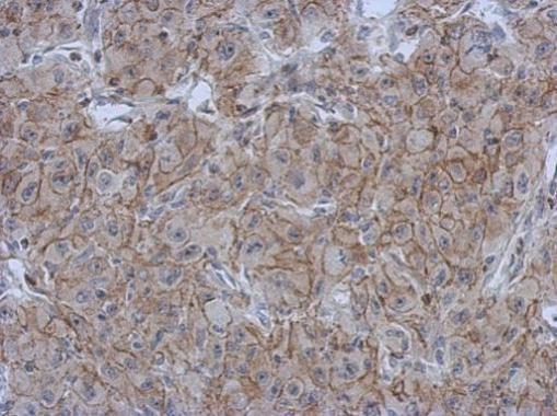
- Immunohistochemical analysis of paraffin-embedded Mahlarvu xenograft, using N-Cadherin(GTX112734) antibody at 1:500 dilution.
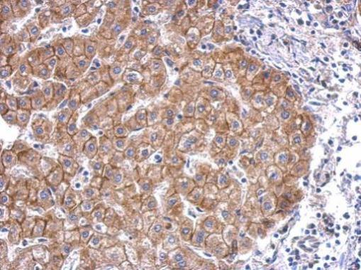
- Submitted by
- GeneTex (provider)
- Main image
- Experimental details
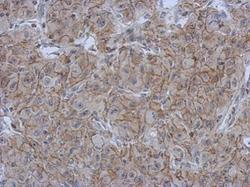
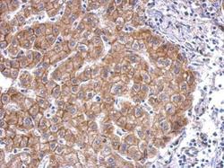
- N-Cadherin antibody [N1N3] detects CDH2 protein at membrane on hepatoma by immunohistochemical analysis. Sample: Paraffin-embedded human hepatoma. N-Cadherin antibody [N1N3] (GTX112734) dilution: 1:500.