 Explore
 Validate
 Learn
Learn Western blot
 Flow cytometry
Antibody data
- Antibody Data
- Antigen structure
- References [1]
- Comments [0]
- Validations
- Western blot [3]
- Immunocytochemistry [1]
- Immunohistochemistry [8]
Submit
Validation data
Reference
Comment
Report error
- Product number
- UM500023 - Provider product page

- Provider
- OriGene
- Proper citation
- OriGene Cat#UM500023, RRID:AB_2629038
- Product name
- CDH2 mouse monoclonal antibody,clone UMAB23
- Antibody type
- Monoclonal
- Description
- CDH2 mouse monoclonal antibody,clone UMAB23
- Host
- Mouse
- Conjugate
- Unconjugated
- Epitope
- CDH2
- Isotype
- IgG
- Antibody clone number
- UMAB23
- Vial size
- 100 µl
- Concentration
- 1.1mg/ml
Submitted references Macrophages significantly enhance wound healing in a vascularized skin model.
Kreimendahl F, Marquardt Y, Apel C, Bartneck M, Zwadlo-Klarwasser G, Hepp J, Jockenhoevel S, Baron JM
Journal of biomedical materials research. Part A 2019 Jun;107(6):1340-1350
Journal of biomedical materials research. Part A 2019 Jun;107(6):1340-1350
No comments: Submit comment
Supportive validation
- Submitted by
- OriGene (provider)
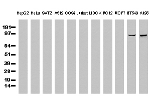
- Main image

- Experimental details
- Western Blot analysis of extracts (35ug) from 11 different cell lines by using anti-CDH2 monoclonal antibody (Clone UMAB23)
- Validation comment
- WB
- Submitted by
- OriGene (provider)
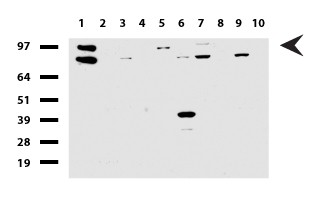
- Main image

- Experimental details
- Western blot of human tissue lysates (15ug) from 10 different tissues (1: Testis, 2: Omentum, 3: Uterus, 4: Breast, 5: Brain, 6: Liver, 7: Ovary, 8: Thyroid, 9: Colon, 10: Spleen ). Diluation: 1:500.
- Validation comment
- WB
- Submitted by
- OriGene (provider)
- Main image

- Experimental details
- Western blot of mouse tissue lysates (20ug) from Brain. Primary antibody diluation: 1:500. Secondary antibody dilution: Mouse TrueBlot? Ultra (1:1000).
- Validation comment
- WB

Supportive validation
- Submitted by
- OriGene (provider)
- Main image
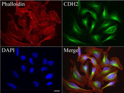
- Experimental details
- Immunofluorescent staining of HeLa cells using anti-CDH2 mouse monoclonal antibody (UM500023, green, 1:50). Actin filaments were labeled with Alexa Fluor? 594 Phalloidin (red), and nuclear with DAPI (blue). Scale bar, 20?m.
- Validation comment
- IF
Supportive validation
- Submitted by
- OriGene (provider)
- Main image

- Experimental details
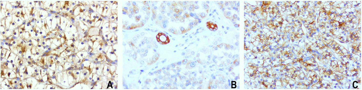
- Immunohistochemical staining of paraffin-embedded of 3 human carcinoma of kidney using anti-CDH2 clone UMAB23 mouse monoclonal antibody at 1:400 dilution of 1.0mg/mL using Polink2 Broad HRP DAB detection kit. UM500023 requires heat-induced epitope retrieval with Citrate pH6.0 in a pressure chamber/cooker high 3min. The composit image of 3 human carcinoma of the kidney all show strong membraneous and cytoplasmic staining in the tumor cells.
- Validation comment
- IHC
- Submitted by
- OriGene (provider)
- Main image
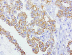
- Experimental details
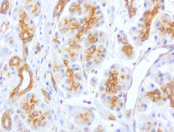
- Immunohistochemical staining of paraffin-embedded of human pancreas using anti-CDH2 clone UMAB23 mouse monoclonal antibody at 1:400 dilution of 1.0mg/mL using Polink2 Broad HRP DAB detection kit. UM500023 requires heat-induced epitope retrieval with Citrate pH6.0 in a pressure chamber/cooker high 3min. The image shows gladular cells of the pancreas membraneous and cytoplasmic staining cells.
- Validation comment
- IHC

- Submitted by
- OriGene (provider)
- Main image

- Experimental details
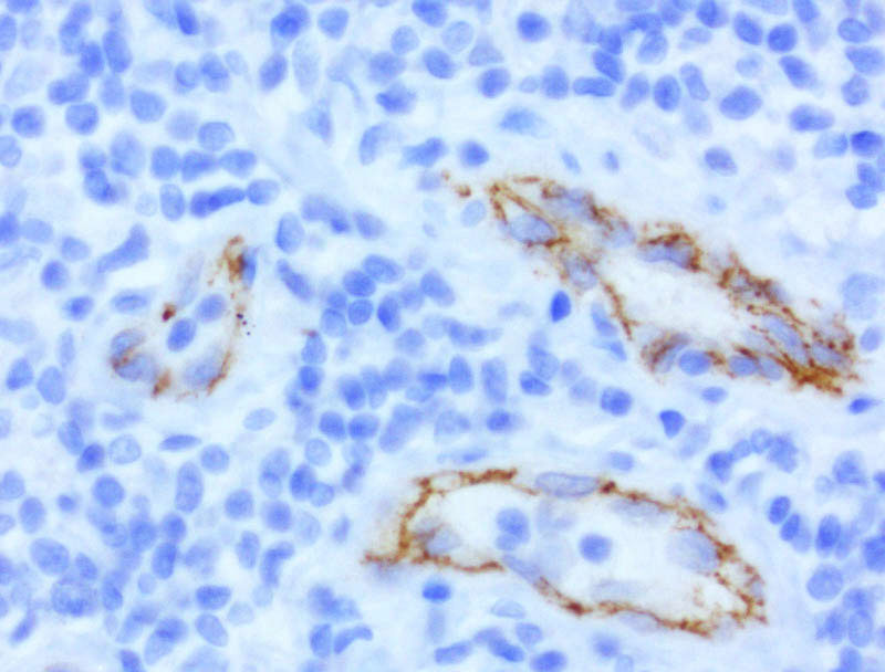
- Immunohistochemical staining of paraffin-embedded of human lymph node using anti-CDH2 clone UMAB23 mouse monoclonal antibody at 1:400 dilution of 1.0mg/mL using Polink2 Broad HRP DAB detection kit. UM500023 requires heat-induced epitope retrieval with Citrate pH6.0 in a pressure chamber/cooker high 3min. The image shows cells of the lymph node are negative. Positive staining seen in the endothelial cells, mostly membraneous with weak cytoplasmic .
- Validation comment
- IHC
- Submitted by
- OriGene (provider)
- Main image
- Experimental details
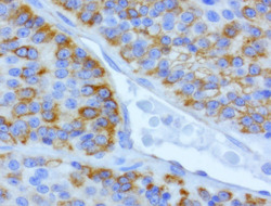
- Immunohistochemical staining of paraffin-embedded of human carcinoma of the lung using anti-CDH2 clone UMAB23 mouse monoclonal antibody at 1:400 dilution of 1.0mg/mL using Polink2 Broad HRP DAB detection kit. UM500023 requires heat-induced epitope retrieval with Citrate pH6.0 in a pressure chamber/cooker high 3min. The image shows the tumor cells with membraneous and cytoplasmic staining.
- Validation comment
- IHC

- Submitted by
- OriGene (provider)
- Main image

- Experimental details
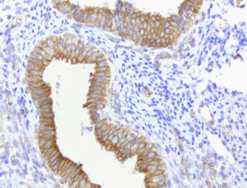
- Immunohistochemical staining of paraffin-embedded of human endometrium using anti-CDH2 clone UMAB23 mouse monoclonal antibody at 1:400 dilution of 1.0mg/mL using Polink2 Broad HRP DAB detection kit. UM500023 requires heat-induced epitope retrieval with Citrate pH6.0 in a pressure chamber/cooker high 3min. The image shows strong membranous and cytoplsamic staining of the endometrial cells.
- Validation comment
- IHC
- Submitted by
- OriGene (provider)
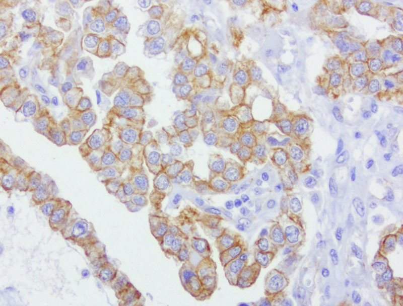
- Main image
- Experimental details
- Immunohistochemical staining of paraffin-embedded of human ovarian carcinoma using anti-CDH2 clone UMAB23 mouse monoclonal antibody at 1:400 dilution of 1.0mg/mL using Polink2 Broad HRP DAB detection kit. UM500023 requires heat-induced epitope retrieval with Citrate pH6.0 in a pressure chamber/cooker high 3min. The image shows strong membranous and cytoplsamic staining of the tumor cells
- Validation comment
- IHC
- Submitted by
- OriGene (provider)
- Main image
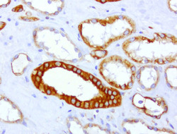
- Experimental details
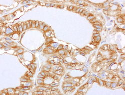
- Immunohistochemical staining of paraffin-embedded of human kidney using anti-CDH2 clone UMAB23 mouse monoclonal antibody at 1:400 dilution of 1.0mg/mL using Polink2 Broad HRP DAB detection kit. UM500023 requires heat-induced epitope retrieval with Citrate pH6.0 in a pressure chamber/cooker high 3min. The image shows strong membranous and cytoplsamic staining in the kidney tubules
- Validation comment
- IHC

- Submitted by
- OriGene (provider)
- Main image
- Experimental details
- Immunohistochemical staining of paraffin-embedded of human thyroid carcinoma using anti-CDH2 clone UMAB23 mouse monoclonal antibody at 1:400 dilution of 1.0mg/mL using Polink2 Broad HRP DAB detection kit. UM500023 requires heat-induced epitope retrieval with Citrate pH6.0 in a pressure chamber/cooker high 3min. The image shows strong membranous and cytoplsamic staining of the tumor cells
- Validation comment
- IHC
