Explore
 Validate
 Learn
Learn Western blot
Antibody data
- Antibody Data
- Antigen structure
- References [1]
- Comments [0]
- Validations
- Western blot [3]
- Immunocytochemistry [3]
- Flow cytometry [1]
Submit
Validation data
Reference
Comment
Report error
- Product number
- 700177 - Provider product page

- Provider
- Invitrogen Antibodies
- Product name
- Phospho-Zap-70 (Tyr315, Tyr319) Recombinant Rabbit Monoclonal Antibody (4H16L19)
- Antibody type
- Monoclonal
- Antigen
- Synthetic peptide
- Description
- This antibody is predicted to react with primate, bovine and Xenopus based on sequence homology.
- Antibody clone number
- 4H16L19
- Concentration
- 0.5 mg/mL
Submitted references Nanoscale kinetic segregation of TCR and CD45 in engaged microvilli facilitates early T cell activation.
Razvag Y, Neve-Oz Y, Sajman J, Reches M, Sherman E
Nature communications 2018 Feb 21;9(1):732
Nature communications 2018 Feb 21;9(1):732
No comments: Submit comment
Supportive validation
- Submitted by
- Invitrogen Antibodies (provider)
- Main image

- Experimental details
- Western blot analysis of Phospho-Zap70 pTyr315/319 in serum-starved, H2O2-treated Jurkat lysate using a Phospho-Zap70 pTyr315/319 recombinant rabbit monoclonal antibody (Product # 700177) at a dilution of 1 µg/mL.
- Submitted by
- Invitrogen Antibodies (provider)
- Main image
- Experimental details
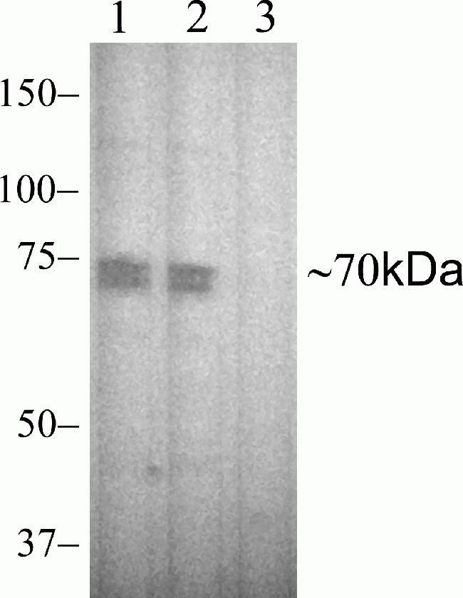
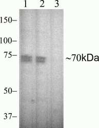
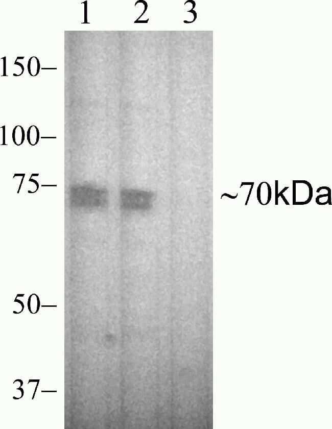
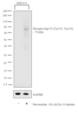
- Western blot analysis was performed on whole cell extracts (30 µg lysate) of MOLT-4 (Lane 1) and MOLT-4 treated with Pervanadate (100uM for 10 minutes) (Lane 2). The blot was probed with Anti-Phospho-Zap-70 (Tyr315, Tyr319) Polyclonal Antibody (Product # 700177, 1:1000 dilution) and detected by chemiluminescence using Goat anti-Rabbit IgG (H+L) Superclonal™ Secondary Antibody, HRP conjugate (Product # A27036, 0.25 µg/ml, 1:4000 dilution). A 70 kDa band corresponding to Phospho-Zap-70 (Tyr315, Tyr319) was induced upon Pervanadate treatment of MOLT-4 cells.

Supportive validation
- Submitted by
- Invitrogen Antibodies (provider)
- Main image

- Experimental details
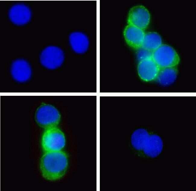
- Immunofluorescent analysis of Phospho-Zap70 pTyr315/319 in Jurkat cells treated with H2O2 (top right) or untreated (top left) using a Phospho-Zap70 pTyr315/319 recombinant rabbit monoclonal antibody (Product # 700177) at a dilution of 2 µg/mL followed by detection using an Alexa Fluor 488-conjugated goat anti-rabbit secondary antibody at a dilution of 1:1000 and nuclei stained using Hoescht (blue). Pre-incubation of treated cells with phosphopeptide immunogen decreased signal (bottom right) while pre-incubation with non-phosphopeptide did not (bottom left).

- Submitted by
- Invitrogen Antibodies (provider)
- Main image

- Experimental details
- Immunofluorescent analysis of Phospho-Zap70 pTyr315/319 in Jurkat cells treated with H2O2 (top right) or untreated (top left) using a Phospho-Zap70 pTyr315/319 recombinant rabbit monoclonal antibody (Product # 700177) at a dilution of 2 µg/mL followed by detection using an Alexa Fluor 488-conjugated goat anti-rabbit secondary antibody at a dilution of 1:1000 and nuclei stained using Hoescht (blue). Pre-incubation of treated cells with phosphopeptide immunogen decreased signal (bottom right) while pre-incubation with non-phosphopeptide did not (bottom left).
- Submitted by
- Invitrogen Antibodies (provider)
- Main image

- Experimental details
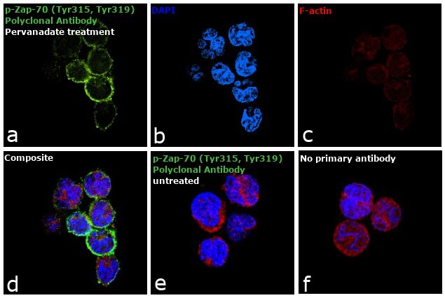
- Immunofluorescence analysis of Phospho-ZAP70 (Tyr 315, 319) was performed using 70% confluent log phase MOLT-4 cells untreated or treated with 100 µM Pervanadate for 10 minutes. The cells were fixed with 4% paraformaldehyde for 10 minutes, permeabilized with 0.1% Triton™ X-100 for 15 minutes, and blocked with 1% BSA for 1 hour at room temperature. The cells were labeled with Anti-Phospho-Zap-70 (Tyr315, Tyr319) Antibody (4H16L19), Recombinant Rabbit Monoclonal (Product # 700177) at 1:200 dilution in 0.1% BSA, incubated at 4 degree Celsius overnight and then labeled with Goat anti-Rabbit IgG (H+L) Superclonal™ Secondary Antibody, Alexa Fluor® 488 conjugate (Product # A27034) at a dilution of 1:2000 for 45 minutes at room temperature (Panel a: green). Nuclei (Panel b: blue) were stained with ProLong™ Diamond Antifade Mountant with DAPI (Product # P36962). F-actin (Panel c: red) was stained with Rhodamine Phalloidin (Product # R415, 1:300). Panel d represents the merged image. Panel e represents untreated control cells while Panel f represents control cells with no primary antibody to assess background. The images were captured at 60X magnification.
Supportive validation
- Submitted by
- Invitrogen Antibodies (provider)
- Main image
- Experimental details
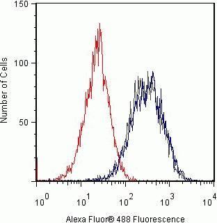
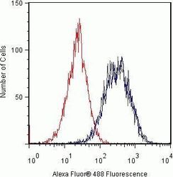
- Flow cytometry analysis of Phospho-Zap70 pTyr315/319 in Jurkat cells using a Phospho-Zap70 pTyr315/319 recombinant rabbit monoclonal antibody (Product # 700177) at a dilution of 2ug. Cells were fixed and permeabilized using FIX & PERM (Product # GAS004) reagent, and detection was performed using an Alexa Fluor 488 goat anti-rabbit IgG (gray) compared to samples pre-incubated with the phosphopeptide (red) and pre-incubated with a non-phospho peptide (blue). Incubation with the phospho peptide decreased the signal while the non-phosphopeptide had no effect.