Explore
 Validate
 Learn
LearnMA1-12710
antibody from Invitrogen Antibodies
Targeting: SNAP25
bA416N4.2, dJ1068F16.2, RIC-4, RIC4, SEC9, SNAP, SNAP-25
 Western blot
 Immunocytochemistry
Antibody data
- Antibody Data
- Antigen structure
- References [0]
- Comments [0]
- Validations
- Immunocytochemistry [1]
Submit
Validation data
Reference
Comment
Report error
- Product number
- MA1-12710 - Provider product page

- Provider
- Invitrogen Antibodies
- Product name
- SNAP25 Monoclonal Antibody (S-7B8)
- Antibody type
- Monoclonal
- Antigen
- Other
- Description
- A suggested positive control for this product is cerebellum tissue lysate.
- Reactivity
- Human, Mouse, Rat, Bovine, Chicken/Avian, Simian
- Host
- Mouse
- Isotype
- IgG
- Antibody clone number
- S-7B8
- Vial size
- 100 µg
- Concentration
- 1 mg/mL
- Storage
- -20° C, Avoid Freeze/Thaw Cycles
No comments: Submit comment
Supportive validation
- Submitted by
- Invitrogen Antibodies (provider)
- Main image
- Experimental details
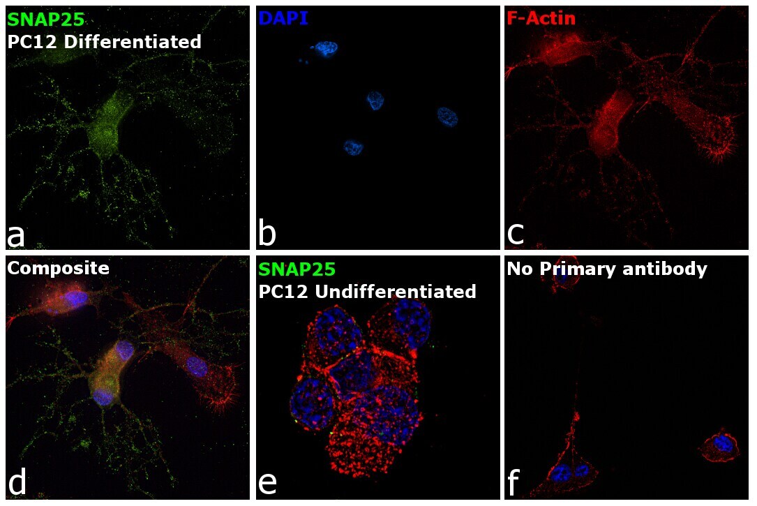
- Immunofluorescence analysis of Synaptosomal-associated protein 25 was performed using 70% confluent log phase PC-12 cells. The cells were fixed with 4% paraformaldehyde for 10 minutes, permeabilized with 0.1% Triton™ X-100 for 15 minutes, and blocked with 2% BSA for 45 minutes at room temperature. The cells were labeled with SNAP25 Monoclonal Antibody (S-7B8) (Product # MA1-12710) at 1:100 dilution in 0.1% BSA, incubated at 4 degree celsius overnight and then labeled with Goat anti-Mouse IgG (H+L) Superclonal™ Recombinant Secondary Antibody, Alexa Fluor® 488 conjugate (Product # A28175), (1:2000 dilution), for 45 minutes at room temperature (Panel a: Green). Nuclei (Panel b: Blue) were stained with ProLong™ Diamond Antifade Mountant with DAPI (Product # P36962). F-actin (Panel c: Red) was stained with Rhodamine Phalloidin (Product # R415, 1:300). Panel d represents the merged image showing enhanced cytoplasmic and membrane localization of SNAP-25 in PC-12 cells differentiated to neuronal phenotype with NGF (200 ng/mL 5 days). Panel e represents the undifferentiated PC-12 cells. Panel f represents control cells with no primary antibody to assess background. The images were captured at 60X magnification.