Explore
 Validate
 Learn
Learn Western blot
Antibody data
- Antibody Data
- Antigen structure
- References [1]
- Comments [0]
- Validations
- Western blot [9]
- Immunocytochemistry [2]
- Immunohistochemistry [3]
- Other assay [1]
Submit
Validation data
Reference
Comment
Report error
- Product number
- PA5-27262 - Provider product page

- Provider
- Invitrogen Antibodies
- Product name
- Syk Polyclonal Antibody
- Antibody type
- Polyclonal
- Antigen
- Recombinant protein fragment
- Description
- Recommended positive controls: A431, Raji, mouse BMDM, mouse spleen.
- Concentration
- 0.32 mg/mL
Submitted references Blood flow guides sequential support of neutrophil arrest and diapedesis by PILR-β1 and PILR-α.
Li YT, Goswami D, Follmer M, Artz A, Pacheco-Blanco M, Vestweber D
eLife 2019 Aug 6;8
eLife 2019 Aug 6;8
No comments: Submit comment
Supportive validation
- Submitted by
- Invitrogen Antibodies (provider)
- Main image

- Experimental details
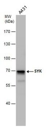
- Western blot analysis of SYK using Whole cell extracts (30 µg). Samples were loaded onto a 7.5% SDS-PAGE gel and probed with a SYK polyclonal antibody (Product # PA5-27262) at a dilution of 1:2000.
- Submitted by
- Invitrogen Antibodies (provider)
- Main image
- Experimental details
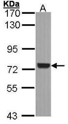
- Western blot analysis of SYK using 30 µg of A431 lysate. Samples were loaded onto a 7.5% SDS-PAGE gel and probed with a SYK polyclonal antibody (Product # PA5-27262) at a dilution of 1:1000.

- Submitted by
- Invitrogen Antibodies (provider)
- Main image
- Experimental details
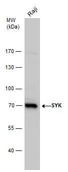
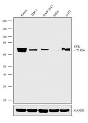
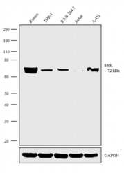
- Western blot analysis was performed on whole cell extracts (30 µg lysate) of Ramos (Lane 1), THP-1 (Lane 2), RAW 264.7 (Lane 3), Jurkat (Lane 4) and A-431 (Lane 5). The blot was probed with Anti-Syk Polyclonal Antibody (Product # PA5-27262, 1:500 dilution) and detected by chemiluminescence using Goat anti-Rabbit IgG (H+L) Superclonal™ Secondary Antibody, HRP conjugate (Product # A27036, 0.25 µg/mL, 1:4000 dilution). A 72 kDa band corresponding to Syk was observed in relevant cell lines.

- Submitted by
- Invitrogen Antibodies (provider)
- Main image
- Experimental details
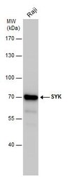
- Western Blot analysis of Syk was performed by separating 30 µg of whole cell extracts by 7.5% SDS-PAGE. Proteins were transferred to a membrane and probed with a Syk Polyclonal Antibody (Product # PA5-27262) at a dilution of 1:2000. The HRP-conjugated anti-rabbit IgG antibody was used to detect the primary antibody.
- Submitted by
- Invitrogen Antibodies (provider)
- Main image
- Experimental details
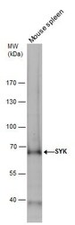
- SYK antibody detects SYK protein by western blot analysis. Mouse tissue extracts (50 µg) was separated by 7.5% SDS-PAGE, and the membrane was blotted with SYK antibody Syk Polyclonal Antibody (Product # PA5-27262) diluted by 1:1,000. The HRP-conjugated anti-rabbit IgG antibody was used to detect the primary antibody.

- Submitted by
- Invitrogen Antibodies (provider)
- Main image
- Experimental details
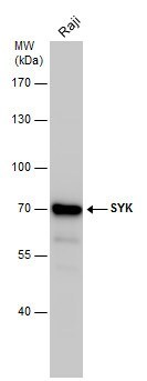
- Western Blot analysis of Syk was performed by separating 30 µg of whole cell extracts by 7.5% SDS-PAGE. Proteins were transferred to a membrane and probed with a Syk Polyclonal Antibody (Product # PA5-27262) at a dilution of 1:2000. The HRP-conjugated anti-rabbit IgG antibody was used to detect the primary antibody.

- Submitted by
- Invitrogen Antibodies (provider)
- Main image
- Experimental details
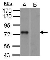
- Syk Polyclonal Antibody detects SYK protein by western blot analysis. A. 30 µg mouse BMDM (bone marrow-derived macrophage) cells. B. 30 µg mouse Syk null cells.10% SDS-PAGE. Syk Polyclonal Antibody (Product # PA5-27262) dilution: 1:1,000. The HRP-conjugated anti-rabbit IgG antibody was used to detect the primary antibody.

- Submitted by
- Invitrogen Antibodies (provider)
- Main image

- Experimental details
- Knockout of SYK was achieved by CRISPR-Cas9 genome editing using LentiArray™ Lentiviral sgRNA (Product # A32042, AssayID CRISPR819133_LV) and LentiArray Cas9 Lentivirus (Product # A32064). Western blot analysis of SYK was performed by loading 30 µg of A-431 wild type (Lane 1), A-431 CAS9 (Lane 2), A-431 SYK KO (Lane 3) whole cell extracts. The blot was probed with Anti-Syk Polyclonal Antibody (Product # PA5-27262) using dilution and Goat anti-Rabbit IgG (H+L), Superclonal™ Recombinant Secondary Antibody, HRP (Product # A27036). Loss of signal upon CRISPR mediated knockout (KO) using the LentiArray™ CRISPR product line confirms that antibody is specific to SYK.

Supportive validation
- Submitted by
- Invitrogen Antibodies (provider)
- Main image
- Experimental details
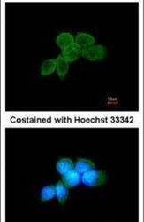
- Immunofluorescent analysis of SYK in methanol-fixed A431 cells using a SYK polyclonal antibody (Product # PA5-27262) at a 1:200 dilution.

- Submitted by
- Invitrogen Antibodies (provider)
- Main image

- Experimental details
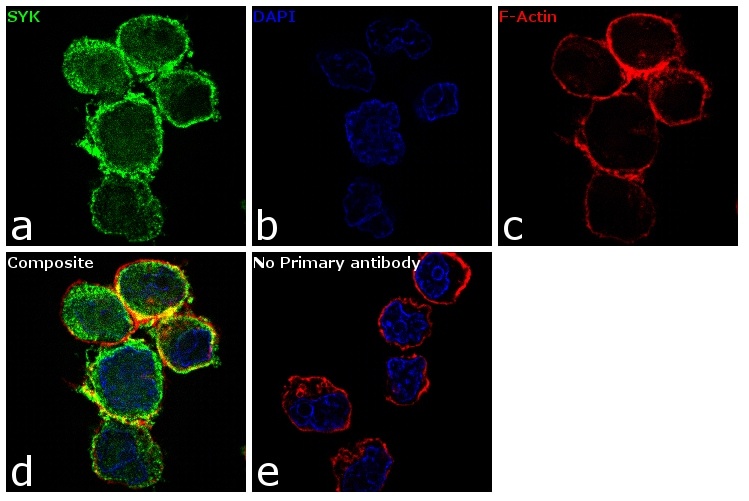
- Immunofluorescence analysis of SYK was performed using 70% confluent log phase Ramos cells. The cells were fixed with 4% paraformaldehyde for 10 minutes, permeabilized with 0.1% Triton™ X-100 for 15 minutes, and blocked with 1% BSA for 1 hour at room temperature. The cells were labeled with SYK Rabbit Polyclonal Antibody(Product # PA5-27262) at 5 µg/mL in 0.1% BSA, incubated at 4 degree Celsius overnight and then labeled with Goat anti-Rabbit IgG (H+L) Superclonal™ Secondary Antibody, Alexa Fluor® 488 conjugate (Product # A27034) at a dilution of 1:2000 for 45 minutes at room temperature (Panel a: green). Nuclei (Panel b: blue) were stained with SlowFade® Gold Antifade Mountant with DAPI (Product # S36938). F-actin (Panel c: red) was stained with Rhodamine Phalloidin (Product # R415, 1:300). Panel d represents the merged image showing cytoplasmic localization. Panel e represents control cells with no primary antibody to assess background. The images were captured at 60X magnification.
Supportive validation
- Submitted by
- Invitrogen Antibodies (provider)
- Main image
- Experimental details
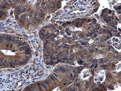
- Immunohistochemistry (Paraffin) analysis of Syk was performed in paraffin-embedded human cervical cancer tissue using Syk Polyclonal Antibody (Product # PA5-27262) at a dilution of 1:500.

- Submitted by
- Invitrogen Antibodies (provider)
- Main image
- Experimental details
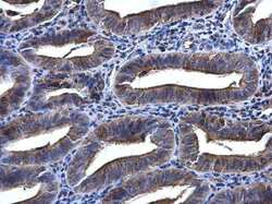
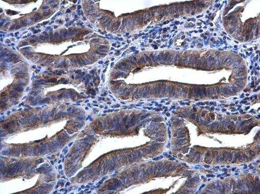
- Immunohistochemistry (Paraffin) analysis of Syk was performed in paraffin-embedded human endometrium tissue using Syk Polyclonal Antibody (Product # PA5-27262) at a dilution of 1:500.
- Submitted by
- Invitrogen Antibodies (provider)
- Main image
- Experimental details
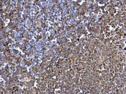
- SYK antibody [N2C2], Internal detects SYK protein at cytosol on rat spleen by immunohistochemical analysis. Sample: Paraffin-embedded rat spleen. SYK antibody [N2C2], Internal (Product # PA5-27262) dilution: 1:500. Antigen Retrieval: EDTA based buffer, pH 8.0, 15 min.

Supportive validation
- Submitted by
- Invitrogen Antibodies (provider)
- Main image

- Experimental details
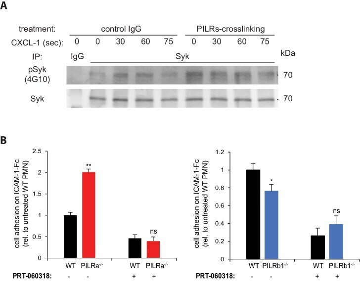
- Figure 3--figure supplement 1. PILRs modulate beta2 integrin activity via Syk. ( A ) PMN were pre-treated with non-crosslinking rabbit IgG or crosslinking polyclonal anti-PILRs for 15 min, and then stimulated with 100 ng/ml CXCL-1 for 0 s (unstimulated), 30 s, 60 s and 75 s, followed by fixation and cell lysis. Syk was immunoprecipitated and analyzed for phospho-Syk (4G10). ( B ) PMNs from WT (black), PILR-alpha -/- (red) or PILR-beta1 -/- (blue) were treated with or without 1 uM Syk-inhibitor PRT-060318 for 30 min. Cells were allowed to adhere to ICAM-1-Fc coated surfaces in the presence of 100 ng/ml CXCL-1 with or without PRT-060318. Adherent cells were counted. n >= 12 for each genotype from at least two experiments. Error bars, SEM. Groups were compared by 2-tailed t-test. ns, not significant, *p