Explore
 Validate
 Learn
Learn Immunocytochemistry
Antibody data
- Antibody Data
- Antigen structure
- References [6]
- Comments [0]
- Validations
- Immunocytochemistry [1]
Submit
Validation data
Reference
Comment
Report error
- Product number
- MA1-12691 - Provider product page

- Provider
- Invitrogen Antibodies
- Product name
- ErbB2 (HER-2) Monoclonal Antibody (N24)
- Antibody type
- Monoclonal
- Antigen
- Other
- Description
- MA1-12691 detects HER-2 in human samples.
- Antibody clone number
- N24
- Concentration
- 1 mg/mL
Submitted references Rapid quantitative profiling of N-glycan by the glycan-labeling method using 3-aminoquinoline/α-cyano-4-hydroxycinnamic acid.
Quantitative assays for the measurement of HER1-HER2 heterodimerization and phosphorylation in cell lines and breast tumors: applications for diagnostics and targeted drug mechanism of action.
BIBW2992, an irreversible EGFR/HER2 inhibitor highly effective in preclinical lung cancer models.
Possible autocrine loop of the epidermal growth factor system in patients with benign prostatic hyperplasia treated with finasteride: a placebo-controlled randomized study.
Possible autocrine loop of the epidermal growth factor system in patients with benign prostatic hyperplasia treated with finasteride: a placebo-controlled randomized study.
Identification of epitope regions recognized by tumor inhibitory and stimulatory anti-ErbB-2 monoclonal antibodies: implications for vaccine design.
Kaneshiro K, Watanabe M, Terasawa K, Uchimura H, Fukuyama Y, Iwamoto S, Sato TA, Shimizu K, Tsujimoto G, Tanaka K
Analytical chemistry 2012 Aug 21;84(16):7146-51
Analytical chemistry 2012 Aug 21;84(16):7146-51
Quantitative assays for the measurement of HER1-HER2 heterodimerization and phosphorylation in cell lines and breast tumors: applications for diagnostics and targeted drug mechanism of action.
DeFazio-Eli L, Strommen K, Dao-Pick T, Parry G, Goodman L, Winslow J
Breast cancer research : BCR 2011 Apr 15;13(2):R44
Breast cancer research : BCR 2011 Apr 15;13(2):R44
BIBW2992, an irreversible EGFR/HER2 inhibitor highly effective in preclinical lung cancer models.
Li D, Ambrogio L, Shimamura T, Kubo S, Takahashi M, Chirieac LR, Padera RF, Shapiro GI, Baum A, Himmelsbach F, Rettig WJ, Meyerson M, Solca F, Greulich H, Wong KK
Oncogene 2008 Aug 7;27(34):4702-11
Oncogene 2008 Aug 7;27(34):4702-11
Possible autocrine loop of the epidermal growth factor system in patients with benign prostatic hyperplasia treated with finasteride: a placebo-controlled randomized study.
Tørring N, Møller-Ernst Jensen K, Lund L, Nielsen JE, Djurhuus JC, Poulsen SS, Nexø E
BJU international 2002 Apr;89(6):583-90
BJU international 2002 Apr;89(6):583-90
Identification of epitope regions recognized by tumor inhibitory and stimulatory anti-ErbB-2 monoclonal antibodies: implications for vaccine design.
Yip YL, Smith G, Koch J, Dübel S, Ward RL
Journal of immunology (Baltimore, Md. : 1950) 2001 Apr 15;166(8):5271-8
Journal of immunology (Baltimore, Md. : 1950) 2001 Apr 15;166(8):5271-8
No comments: Submit comment
Supportive validation
- Submitted by
- Invitrogen Antibodies (provider)
- Main image
- Experimental details
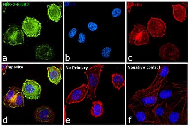
- Immunofluorescence analysis of Her2 was performed using 70% confluent log phase SK-BR-3 cells. The cells were fixed with 4% paraformaldehyde for 10 minutes, permeabilized with 0.1% Triton™ X-100 for 10 minutes, and blocked with 1% BSA for 1 hour at room temperature. The cells were labeled with Her2 Mouse monoclonal Antibody (Product # MA1-12691) at 5 µg/mL in 0.1% BSA and incubated overnight at 4 degree Celsius and then labeled with Goat anti-Mouse IgG (H+L) Superclonal™ Secondary Antibody, Alexa Fluor® 488 conjugate (Product # A28175) at a dilution of 1:2000 for 45 minutes at room temperature (Panel a: green). Nuclei (Panel b: blue) were stained with SlowFade® Gold Antifade Mountant with DAPI (Product # S36938). F-actin (Panel c: red) was stained with Rhodamine Phalloidin (Product # R415, 1:300). Panel d represents the merged image showing membranous and cytoplasmic localization. Panel f represents MDAMB-231 cells as negative controls, showing no Her2 staining. Panel e represents control cells with no primary antibody to assess background. The images were captured at 60X magnification.