 Explore
 Validate
 Learn
Learn Western blot
Antibody data
- Antibody Data
- Antigen structure
- References [0]
- Comments [0]
- Validations
- Western blot [2]
- Immunocytochemistry [2]
- Immunohistochemistry [1]
- Flow cytometry [1]
Submit
Validation data
Reference
Comment
Report error
- Product number
- ABIN2508060 - Provider product page

- Provider
- antibodies-online
- Product name
- anti-V-Erb-B2 erythroblastic Leukemia Viral Oncogene Homolog 2, Neuro/glioblastoma Derived Oncogene Homolog (Avian) (ERBB2) (pTyr877) antibody
- Antibody type
- Polyclonal
- Antigen
- This ERBB2 Antibody is generated from rabbits immunized with a KLH conjugated synthetic phosphopeptide corresponding to amino acid residues surrounding Y877 of human ERBB2.Antigen type: Synthetic Peptide
- Description
- This antibody is purified through a protein A column, followed by peptide affinity purification.
- Reactivity
- Human
- Host
- Rabbit
- Epitope
- pTyr877
- Vial size
- 200 μL
- Storage
- Maintain refrigerated at 2-8°C for up to 6 months. For long term storage store at -20°C in small aliquots to prevent freeze-thaw cycles.
No comments: Submit comment
Supportive validation
- Submitted by
- antibodies-online (provider)
- Main image

- Experimental details
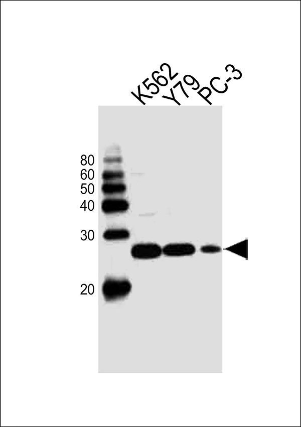
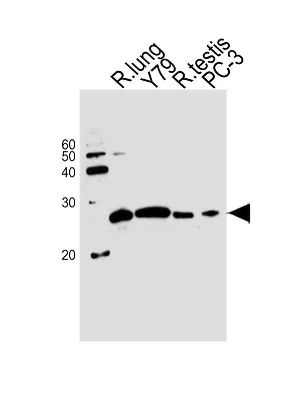
- Western blot analysis of lysates from K562,Y79,PC-3 cell line (from left to right), using GSTP1 Antibody (Center)(Cat. ABIN2509767). ABIN2509767 was diluted at 1:1000 at each lane. A goat anti-rabbit IgG H&L(HRP) at 1:10000 dilution was used as the secondary antibody. Western blot analysis of lysates from rat lung tissue,Y79 cell line,rat testis tissue,PC-3 cell line (from left to right), using GSTP1 Antibody (Center)(Cat. ABIN2509767). ABIN2509767 was diluted at 1:1000 at each lane. A goat anti-rabbit IgG H&L(HRP) at 1:10000 dilution was used as the secondary antibody.Lysates at 20ug per lane. Western blot analysis of lysates from LNCaP cell line,mouse kidney and rat kidney tissue lysate(from left to right),using MME Antibody (Center)(Cat. WA1025). WA1025 was diluted at 1:1000 at each lane. A goat anti-rabbit IgG H&L(HRP) at 1:10000 dilution was used as the secondary antibody. Western blot analysis of lysates from mouse lung tissue lysate,Y79,K562,PC-3 cell line (from left to right), using GSTP1 Antibody (Center)(Cat. ABIN2509767). ABIN2509767 was diluted at 1:1000 at each lane. A goat anti-rabbit IgG H&L(HRP) at 1:10000 dilution was used as the secondary antibody.Lysates at 20ug per lane.
- Submitted by
- antibodies-online (provider)
- Main image

- Experimental details
- Western blot analysis of lysates from K562,Y79,PC-3 cell line (from left to right), using GSTP1 Antibody (Center)(Cat. ABIN2509767). ABIN2509767 was diluted at 1:1000 at each lane. A goat anti-rabbit IgG H&L(HRP) at 1:10000 dilution was used as the secondary antibody. Western blot analysis of lysates from rat lung tissue,Y79 cell line,rat testis tissue,PC-3 cell line (from left to right), using GSTP1 Antibody (Center)(Cat. ABIN2509767). ABIN2509767 was diluted at 1:1000 at each lane. A goat anti-rabbit IgG H&L(HRP) at 1:10000 dilution was used as the secondary antibody.Lysates at 20ug per lane. Western blot analysis of lysates from LNCaP cell line,mouse kidney and rat kidney tissue lysate(from left to right),using MME Antibody (Center)(Cat. WA1025). WA1025 was diluted at 1:1000 at each lane. A goat anti-rabbit IgG H&L(HRP) at 1:10000 dilution was used as the secondary antibody. Western blot analysis of lysates from mouse lung tissue lysate,Y79,K562,PC-3 cell line (from left to right), using GSTP1 Antibody (Center)(Cat. ABIN2509767). ABIN2509767 was diluted at 1:1000 at each lane. A goat anti-rabbit IgG H&L(HRP) at 1:10000 dilution was used as the secondary antibody.Lysates at 20ug per lane.
Supportive validation
- Submitted by
- antibodies-online (provider)
- Main image
- Experimental details
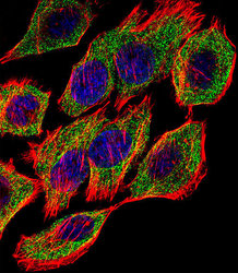
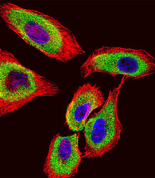
- Image(s): Immunofluorescence
- Submitted by
- antibodies-online (provider)
- Main image
- Experimental details
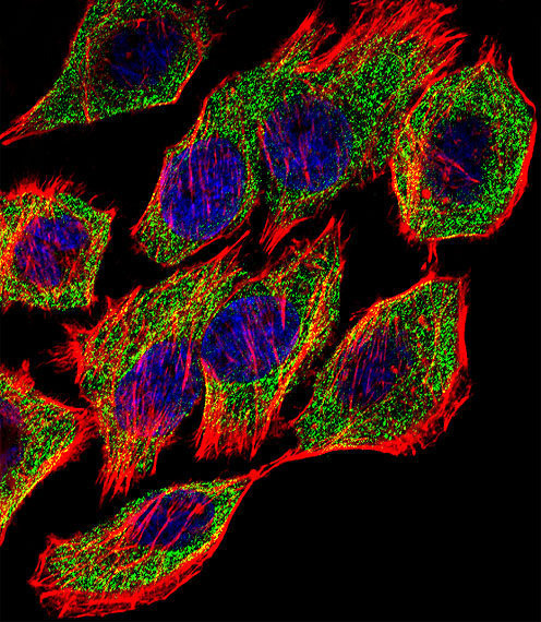
- Image(s): Immunofluorescence

Supportive validation
- Submitted by
- antibodies-online (provider)
- Main image

- Experimental details
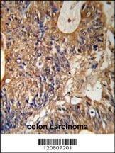
- Image(s): Immunohistochemistry
Supportive validation
- Submitted by
- antibodies-online (provider)
- Main image
- Experimental details
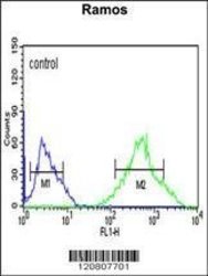
- Image(s): Flow cytometry
