Explore
 Validate
 Learn
Learn Western blot
Antibody data
- Antibody Data
- Antigen structure
- References [2]
- Comments [0]
- Validations
- Western blot [2]
- Immunohistochemistry [1]
Submit
Validation data
Reference
Comment
Report error
- Product number
- AF1768 - Provider product page

- Provider
- R&D Systems
- Product name
- Human Phospho-ErbB2/Her2 (Y1248) Antibody
- Antibody type
- Polyclonal
- Description
- Antigen Affinity-purified. Detects human ErbB2 when phosphorylated at Y1248.
- Reactivity
- Human
- Host
- Rabbit
- Conjugate
- Unconjugated
- Isotype
- IgG
- Vial size
- 50 ug
- Concentration
- LYOPH
- Storage
- Use a manual defrost freezer and avoid repeated freeze-thaw cycles. 12 months from date of receipt, -20 to -70 °C as supplied. 1 month, 2 to 8 °C under sterile conditions after reconstitution. 6 months, -20 to -70 °C under sterile conditions after reconstitution.
Submitted references Modulation of ErbB2 blockade in ErbB2-positive cancers: the role of ErbB2 Mutations and PHLDA1.
Osteoblast-derived factors induce an expression signature that identifies prostate cancer metastasis and hormonal progression.
Li G, Wang X, Hibshoosh H, Jin C, Halmos B
PloS one 2014;9(9):e106349
PloS one 2014;9(9):e106349
Osteoblast-derived factors induce an expression signature that identifies prostate cancer metastasis and hormonal progression.
Wang G, Haile S, Comuzzi B, Tien AH, Wang J, Yong TM, Jelescu-Bodos AE, Blaszczyk N, Vessella RL, Masri BA, Sadar MD
Cancer research 2009 Apr 15;69(8):3433-42
Cancer research 2009 Apr 15;69(8):3433-42
No comments: Submit comment
Supportive validation
- Submitted by
- R&D Systems (provider)
- Main image
- Experimental details
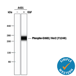
- Detection of Human Phospho-ErbB2/Her2 (Y1248) by Simple WesternTM. Simple Western lane view shows lysates of A431 human epithelial carcinoma cell line untreated (-) or treated (+) with 10 ng/mL Recombinant Human EGF (Catalog # 236-EG) for 5 minutes, loaded at 0.2 mg/mL. A specific band was detected for Phospho-ErbB2/Her2 (Y1248) at approximately 265 kDa (as indicated) using 5 µg/mL of Rabbit Anti-Human Phospho-ErbB2/Her2 (Y1248) Antigen Affinity-purified Polyclonal Antibody (Catalog # AF1768). This experiment was conducted under reducing conditions and using the 66-440 kDa separation system.

- Submitted by
- R&D Systems (provider)
- Main image
- Experimental details
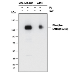
- Detection of Human Phospho-ErbB2/Her2 (Y1248) by Western Blot. Western blot shows lysates of MDA-MB-468 human breast cancer cell line and A431 human epithelial carcinoma cell line untreated (-) or treated (+) with 1 mM Pervanadate (PV) for 10 minutes or with 10 ng/mL Recombinant Human EGF (Catalog # 236-EG) for 5 minutes. PVDF membrane was probed with 0.25 µg/mL of Rabbit Anti-Human Phospho-ErbB2/Her2 (Y1248) Antigen Affinity-purified Polyclonal Antibody (Catalog # AF1768) followed by HRP-conjugated Anti-Rabbit IgG Secondary Antibody (Catalog # HAF008). A specific band was detected for Phospho-ErbB2/Her2 (Y1248) at approximately 170 kDa (as indicated). This experiment was conducted under reducing conditions and using Immunoblot Buffer Group 1.
Supportive validation
- Submitted by
- R&D Systems (provider)
- Main image

- Experimental details
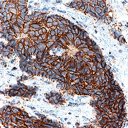
- ErbB2/Her2 in Human Breast Cancer Tissue. ErbB2/Her2 was detected in immersion fixed paraffin-embedded sections of human breast cancer tissue using Rabbit Anti-Human Phospho-ErbB2/Her2 (Y1248) Antigen Affinity-purified Polyclonal Antibody (Catalog # AF1768) at 0.3 µg/mL for 1 hour at room temperature followed by incubation with the Anti-Rabbit IgG VisUCyte™ HRP Polymer Antibody (Catalog # VC003). Before incubation with the primary antibody, tissue was subjected to heat-induced epitope retrieval using Antigen Retrieval Reagent-Basic (Catalog # CTS013). Tissue was stained using DAB (brown) and counterstained with hematoxylin (blue). Specific staining was localized to plasma membrane. View our protocol for IHC Staining with VisUCyte HRP Polymer Detection Reagents.