 Explore
 Validate
 Learn
Learn Western blot
Antibody data
- Antibody Data
- Antigen structure
- References [1]
- Comments [0]
- Validations
- Western blot [4]
- Immunocytochemistry [1]
- Immunohistochemistry [5]
Submit
Validation data
Reference
Comment
Report error
- Product number
- HPA002192 - Provider product page

- Provider
- Atlas Antibodies
- Proper citation
- Atlas Antibodies Cat#HPA002192, RRID:AB_1079695
- Product name
- Anti-LONP1
- Antibody type
- Polyclonal
- Reactivity
- Human, Mouse, Rat
- Host
- Rabbit
- Conjugate
- Unconjugated
- Antigen sequence
VEEKIKQTHRKYLLQEQLKIIKKELGLEKDDKDAI
EEKFRERLKELVVPKHVMDVVDEELSKLGLLDNHS
SEFNVTRNYLDWLTSIPWGKYSNENLDLARAQAVL
EEDHYGMEDVKKRILEFIAVSQLRGSTQGKILCFY
GP- Isotype
- IgG
- Vial size
- 100 µl
- Storage
- Store at +4°C for short term storage. Long time storage is recommended at -20°C.
Submitted references Proteomics Analysis Reveals Novel Components in the Detergent-Insoluble Subproteome in Alzheimer’s Disease
Gozal Y, Duong D, Gearing M, Cheng D, Hanfelt J, Funderburk C, Peng J, Lah J, Levey A
Journal of Proteome Research 2009 November;8(11):5069-5079
Journal of Proteome Research 2009 November;8(11):5069-5079
No comments: Submit comment
Supportive validation
Supportive validation
- Submitted by
- Atlas Antibodies (provider)
- Enhanced method
- Genetic validation
- Main image

- Experimental details
- Western blot analysis in A-431 cells transfected with control siRNA, target specific siRNA probe #1 and #2, using Anti-LONP1 antibody. Remaining relative intensity is presented. Loading control: Anti-GAPDH.

- Submitted by
- Atlas Antibodies (provider)
- Enhanced method
- Independent antibody validation
- Main image

- Experimental details
- Western blot analysis using Anti-LONP1 antibody HPA002192 (A) shows similar pattern to independent antibody HPA002034 (B).

Supportive validation
- Submitted by
- Atlas Antibodies (provider)
- Main image
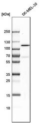
- Experimental details
- Western blot analysis in human cell line SK-MEL-30.

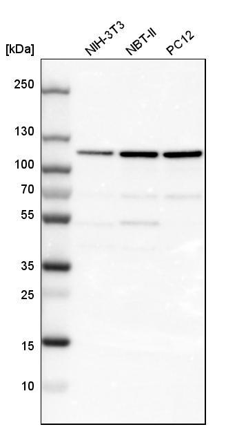
- Submitted by
- Atlas Antibodies (provider)
- Main image
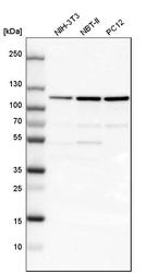
- Experimental details
- Western blot analysis in mouse cell line NIH-3T3, rat cell line NBT-II and rat cell line pC12.
Supportive validation
- Submitted by
- Atlas Antibodies (provider)
- Main image
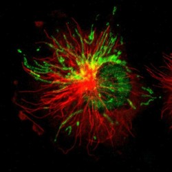
- Experimental details
- Immunofluorescent staining of human cell line U-251 MG shows localization to nucleoplasm & mitochondria.
- Sample type
- HUMAN

Supportive validation
- Submitted by
- Atlas Antibodies (provider)
- Main image

- Experimental details
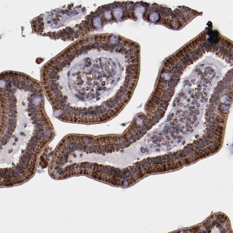
- Immunohistochemical staining of human duodenum shows strong cytoplasmic positivity in glandular cells.
- Submitted by
- Atlas Antibodies (provider)
- Main image

- Experimental details
- Immunohistochemical staining of human fallopian tube shows granular cytoplasmic positivity in glandular cells.
- Sample type
- HUMAN

- Submitted by
- Atlas Antibodies (provider)
- Main image
- Experimental details
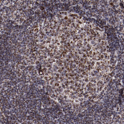
- Immunohistochemical staining of human tonsil shows granular cytoplasmic positivity.
- Sample type
- HUMAN

- Submitted by
- Atlas Antibodies (provider)
- Main image
- Experimental details
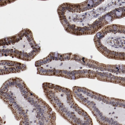
- Immunohistochemical staining of human duodenum shows granular cytoplasmic positivity in glandular cells.
- Sample type
- HUMAN

- Submitted by
- Atlas Antibodies (provider)
- Main image

- Experimental details
- Immunohistochemical staining of human pancreas shows granular cytoplasmic positivity.
- Sample type
- HUMAN
