 Explore
 Validate
 Learn
Learn Western blot
Antibody data
- Antibody Data
- Antigen structure
- References [2]
- Comments [0]
- Validations
- Western blot [1]
- Immunocytochemistry [1]
- Immunohistochemistry [2]
- Other assay [2]
Submit
Validation data
Reference
Comment
Report error
- Product number
- MA1-06908 - Provider product page

- Provider
- Invitrogen Antibodies
- Product name
- Vimentin Monoclonal Antibody (RV203)
- Antibody type
- Monoclonal
- Antigen
- Other
- Description
- MA1-06908 detects vimentin in human, rat, goat, canine, hamster and swine samples.
- Antibody clone number
- RV203
- Concentration
- 1 mg/mL
Submitted references Functional Expression of Transient Receptor Potential and Piezo1 Channels in Cultured Interstitial Cells of Human-Bladder Lamina Propria.
Cancer associated fibroblasts transfer lipids and proteins to cancer cells through cargo vesicles supporting tumor growth.
Zhao M, Chen Z, Liu L, Ding N, Wen J, Liu J, Wang W, Ge N, Zu S, Song W, Chen G, Zhang X
Frontiers in physiology 2021;12:762847
Frontiers in physiology 2021;12:762847
Cancer associated fibroblasts transfer lipids and proteins to cancer cells through cargo vesicles supporting tumor growth.
Santi A, Caselli A, Ranaldi F, Paoli P, Mugnaioni C, Michelucci E, Cirri P
Biochimica et biophysica acta 2015 Dec;1853(12):3211-23
Biochimica et biophysica acta 2015 Dec;1853(12):3211-23
No comments: Submit comment
Supportive validation
- Submitted by
- Invitrogen Antibodies (provider)
- Main image

- Experimental details
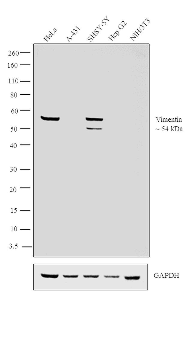
- Western blot analysis was performed on Whole cell extracts (30 µg lysate) of HeLa (Lane 1), A-431 (Lane 2), SHSY-5Y (Lane 3), Hep G2 (Lane 4) and NIH/3T3 (Lane 5). The blot was probed with Anti-Vimentin Monoclonal Antibody (Product # MA1-06908, 1:2000 dilution) and detected by chemiluminescence using Goat anti-Mouse IgG (H+L) Superclonal™ Secondary Antibody, HRP conjugate (Product # A28177, 0.25 µg/ml, 1:4000 dilution). A 54 kDa band corresponding to Vimentin was observed across all the human cell lines positive for Vimentin (Lanes 1 and 3) but not mouse cell line (Lane 5), while this band was absent in the cell lines which do not express Vimentin protein (Lanes 2 and 4).
Supportive validation
- Submitted by
- Invitrogen Antibodies (provider)
- Main image

- Experimental details
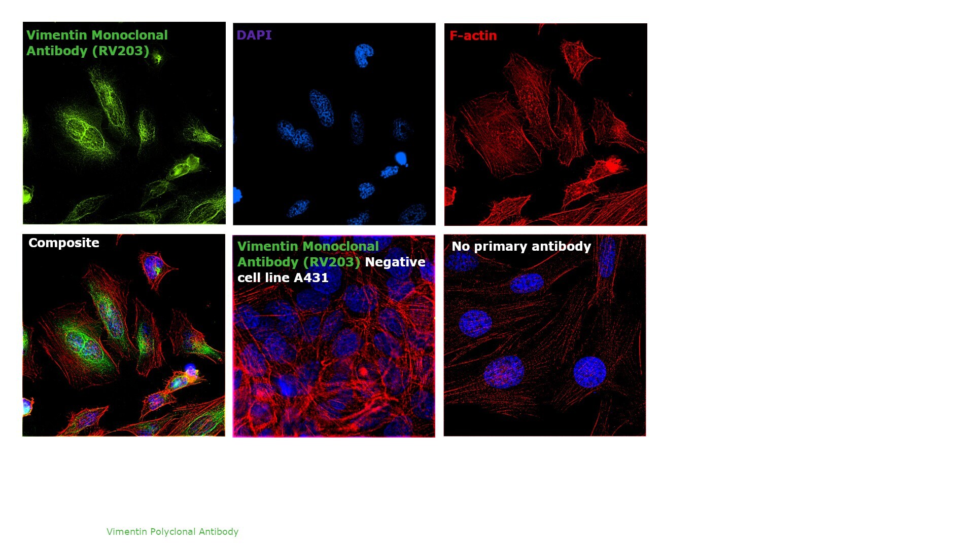
- Immunofluorescence analysis of Vimentin was performed using 70% confluent log phase HeLa cells. The cells were fixed with 4% paraformaldehyde for 10 minutes, permeabilized with 0.1% Triton™ X-100 for 10 minutes, and blocked with 1% BSA for 1 hour at room temperature. The cells were labeled with Vimentin Mouse Monoclonal Antibody (Product # MA1-06908) at 5 µg/mL in 0.1% BSA and incubated overnight at 4 degree Celsius and then labeled with Goat anti-Mouse IgG (H+L) Superclonal™ Secondary Antibody, Alexa Fluor® 488 conjugate (Product # A28175) at a dilution of 1:2000 for 45 minutes at room temperature (Panel a: green). Nuclei (Panel b: blue) were stained with SlowFade® Gold Antifade Mountant with DAPI (Product # S36938). F-actin (Panel c: red) was stained with Rhodamine Phalloidin (Product # R415, 1:300). Panel d represents the merged image showing cytoplasmic, cytoskeletal and nuclear localization. Panel e represents negative control, A-431 cells. Panel f represents control cells with no primary antibody to assess background. The images were captured at 60X magnification.
Supportive validation
- Submitted by
- Invitrogen Antibodies (provider)
- Main image
- Experimental details
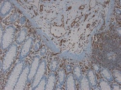
- Immunohistochemistry on frozen section of human colon showing positive staining in connective tissue cells and no reactivity in epithelial cells stained with Vimentin monoclonal antibody (Product # MA1-06908).

- Submitted by
- Invitrogen Antibodies (provider)
- Main image
- Experimental details
- Immunohistochemistry on paraffin section of human colon stained with Vimentin monoclonal antibody (Product # MA1-06908).

Supportive validation
- Submitted by
- Invitrogen Antibodies (provider)
- Main image
- Experimental details
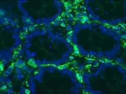
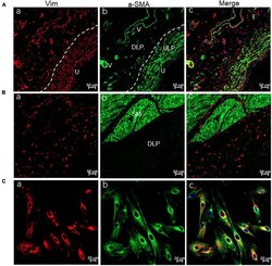
- FIGURE 1 Confocal immunofluorescence for Vim (red) and alpha-SMA (green) showing ULP-ICs and cultured ICs having the phenotype of Vim+ alpha-SMA+. (A,B) Immunofluorescence of the bladder wall showing Vim+ (red) ICs distributed in the ULP [ (Aa) , densely packed cells immediately beneath the urothelium], DLP [ (Ba) , loosely distributed between the ULP and detrusor] as well within or between the detrusor muscle (Ba) . Vim+ staining is also seen in endothelial cells of blood vessels (V). alpha-SMA+ (green) staining is present on ULP-ICs (Ab) and the detrusor muscle [SM, (Bb) ] but not on DLP-ICs or detrusor-ICs (Bb) . Perivascular smooth muscle also expresses alpha-SMA. Co-expression of Vim and alpha-SMA is shown in merged images (Ac and Bc) . (Ca-Cc) Immunofluorescence of Vim and alpha-SMA in cultured LP-ICs showing most of the Vim+ ICs are alpha-SMA+. DLP: deep lamina propria; ULP: upper lamina propria; U: urothelium; V: vessels; SM: smooth muscle. Dashed line indicates the transition between the ULP and DLP.

- Submitted by
- Invitrogen Antibodies (provider)
- Main image
- Experimental details
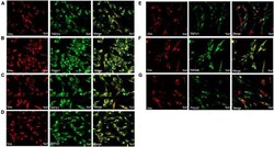
- FIGURE 3 Immunofluorescence for Vim (red) and TRP or Piezo channels (green) in cultured LP-ICs. Double staining reveals the protein expression of TRPV4 (A) , piezo1 (B) , TRPA1 (C) , TRPV2 (D) , TRPV1 (E) , TRPM8 (F) and Piezo2 (G) in most of the Vim+ LP-ICs. The nucleus marker (DAPI) is stained in blue.
