Explore
 Validate
 Learn
Learn Western blot
Antibody data
- Antibody Data
- Antigen structure
- References [0]
- Comments [0]
- Validations
- Western blot [3]
- Immunocytochemistry [2]
- Immunohistochemistry [4]
Submit
Validation data
Reference
Comment
Report error
- Product number
- MA5-26272 - Provider product page

- Provider
- Invitrogen Antibodies
- Product name
- Arginase 2 Monoclonal Antibody (OTI3G5)
- Antibody type
- Monoclonal
- Antigen
- Recombinant full-length protein
- Reactivity
- Human, Mouse, Rat, Canine
- Host
- Mouse
- Isotype
- IgG
- Antibody clone number
- OTI3G5
- Vial size
- 100 µL
- Concentration
- 1 mg/mL
- Storage
- -20° C, Avoid Freeze/Thaw Cycles
No comments: Submit comment
Supportive validation
- Submitted by
- Invitrogen Antibodies (provider)
- Main image
- Experimental details
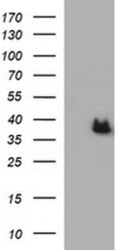
- Western blot analysis of ARG2 in HEK293T cells in untransfected (Left lane) and transfected (Right lane) samples using 5 µg per lane. The samples were separated by SDS-PAGE and probed with ARG2 (Product # MA5-26272) monoclonal antibody.

- Submitted by
- Invitrogen Antibodies (provider)
- Main image
- Experimental details
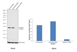
- Knockdown of Arginase 2 was achieved by transfecting LNCaP with Arginase 2 specific siRNA (Silencer® select Product # s1571). Western blot analysis (Fig. a) was performed using Whole cell extracts from the Arginase 2 knockdown cells (lane 3), non-specific scrambled siRNA transfected cells (lane 2) and untransfected cells (lane 1). The blot was probed with Arginase 2 Monoclonal Antibody (Product # MA5-26272, 1:1000 dilution) and Goat anti-Mouse IgG (H+L) Superclonal™ Recombinant Secondary Antibody, HRP (Product # A28177, 0.25µg/ml, 1:4000 dilution). Densitometric analysis of this western blot is shown in histogram (Fig. b). Decrease in signal upon siRNA mediated knock down confirms that antibody is specific to Arginase 2.

- Submitted by
- Invitrogen Antibodies (provider)
- Main image
- Experimental details
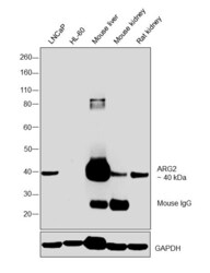
- Western blot was performed using Anti-Arginase 2 Monoclonal Antibody (OTI3G5) (Product # MA5-26272) and a 40 kDa band corresponding to Arginase 2 along with an uncharacterized band at ~90 kDa in Mouse liver and circulating mouse IgG at ~25 kDa was observed in mouse tissues tested were observed. HL-60 showed no expression compared to LNCaP. Whole cell extracts (40 µg lysate) of LNCaP (Lane 1), HL-60 (Lane 2), Mouse liver (Lane 3), Mouse kidney (Lane 4), and Rat kidney (Lane 5) were electrophoresed using Novex® NuPAGE® 4-12 % Bis-Tris gel (Product # NP0321BOX). Resolved proteins were then transferred onto a nitrocellulose membrane (Product # IB23001) by iBlot® 2 Dry Blotting System (Product # IB21001). The blot was probed with the primary antibody (1:2000 dilution) and detected by chemiluminescence with Goat anti-Mouse IgG (H+L), Superclonal™ Recombinant Secondary Antibody, HRP conjugate (Product # A28177, 1:4000 dilution) using the iBright FL 1000 (Product # A32752). Chemiluminescent detection was performed using Novex® ECL Chemiluminescent Substrate Reagent Kit (Product # WP20005).

Supportive validation
- Submitted by
- Invitrogen Antibodies (provider)
- Main image
- Experimental details
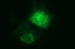
- Immunofluorescent analysis of ARG2 in COS7 cells. Cells were transfected with a plasmid overexpressing ARG2 and probed with a ARG2 monoclonal antibody (Product # MA5-26272).

- Submitted by
- Invitrogen Antibodies (provider)
- Main image

- Experimental details
- Immunofluorescent analysis of ARG2 in COS7 cells. Cells were transfected with a plasmid overexpressing ARG2 and probed with a ARG2 monoclonal antibody (Product # MA5-26272).

Supportive validation
- Submitted by
- Invitrogen Antibodies (provider)
- Main image
- Experimental details
- Immunohistochemistry was performed on paraffin-embedded adenocarcinoma of human colon tissue. To expose target proteins, 10mM citric buffer, pH6.0, 120°C for 3min was used. Following antigen retrieval, tissues were probed with a ARG2 monoclonal antibody (Product # MA5-26272).

- Submitted by
- Invitrogen Antibodies (provider)
- Main image

- Experimental details
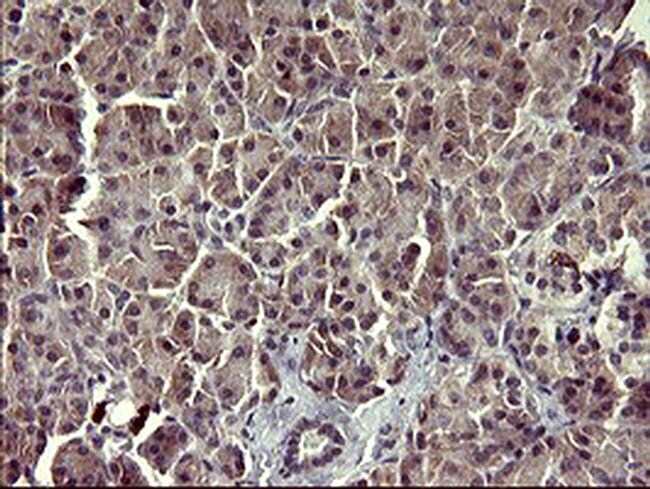
- Immunohistochemistry was performed on paraffin-embedded human pancreas tissue. To expose target proteins, 10mM citric buffer, pH6.0, 120°C for 3min was used. Following antigen retrieval, tissues were probed with a ARG2 monoclonal antibody (Product # MA5-26272).
- Submitted by
- Invitrogen Antibodies (provider)
- Main image
- Experimental details
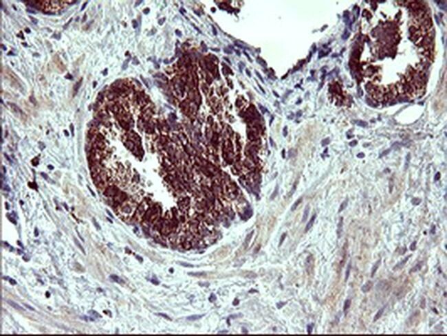
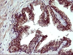
- Immunohistochemistry was performed on paraffin-embedded human prostate tissue. To expose target proteins, 10mM citric buffer, pH6.0, 120°C for 3min was used. Following antigen retrieval, tissues were probed with a ARG2 monoclonal antibody (Product # MA5-26272).

- Submitted by
- Invitrogen Antibodies (provider)
- Main image

- Experimental details
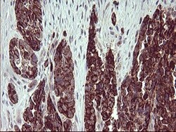
- Immunohistochemistry was performed on paraffin-embedded carcinoma of human prostate tissue. To expose target proteins, 10mM citric buffer, pH6.0, 120°C for 3min was used. Following antigen retrieval, tissues were probed with a ARG2 monoclonal antibody (Product # MA5-26272).