Explore
 Validate
 Learn
Learn Western blot
Antibody data
- Antibody Data
- Antigen structure
- References [1]
- Comments [0]
- Validations
- Western blot [1]
- Immunohistochemistry [1]
Submit
Validation data
Reference
Comment
Report error
- Product number
- AF7514 - Provider product page

- Provider
- Novus Biologicals
- Product name
- Sheep Polyclonal TM4SF1/L6 Antibody
- Antibody type
- Polyclonal
- Description
- Immunogen affinity purified. Detects mouse TM4SF1 in direct ELISAs and Western blots.
- Reactivity
- Mouse
- Host
- Sheep
- Isotype
- IgG
- Vial size
- 100 ug
- Concentration
- LYOPH
- Storage
- Use a manual defrost freezer and avoid repeated freeze-thaw cycles. 12 months from date of receipt, -20 to -70 degreesC as supplied. 1 month, 2 to 8 degreesC under sterile conditions after reconstitution. 6 months, -20 to -70 degreesC under sterile conditions after reconstitution.
Submitted references Regeneration of the lung alveolus by an evolutionarily conserved epithelial progenitor.
Zacharias WJ, Frank DB, Zepp JA, Morley MP, Alkhaleel FA, Kong J, Zhou S, Cantu E, Morrisey EE
Nature 2018 Mar 8;555(7695):251-255
Nature 2018 Mar 8;555(7695):251-255
No comments: Submit comment
Supportive validation
- Submitted by
- Novus Biologicals (provider)
- Main image
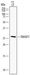
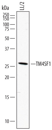
- Experimental details
- Detection of Mouse TM4SF1 by Western Blot. Western blot shows lysates of LL/2 mouse Lewis lung carcinoma cell line. PVDF membrane was probed with 1 µg/mL of Sheep Anti-Mouse TM4SF1 Antigen Affinity-purified Polyclonal Antibody (Catalog # AF7514) followed by HRP-conjugated Anti-Sheep IgG Secondary Antibody (Catalog # HAF016). A specific band was detected for TM4SF1 at approximately 26 kDa (as indicated). This experiment was conducted under reducing conditions and using Immunoblot Buffer Group 1.
Supportive validation
- Submitted by
- Novus Biologicals (provider)
- Main image

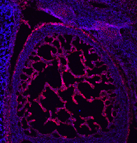
- Experimental details
- TM4SF1 in Mouse Embryo. TM4SF1 was detected in immersion fixed frozen sections of mouse embryo (13 d.p.c.) using Sheep Anti-Mouse TM4SF1 Antigen Affinity-purified Polyclonal Antibody (Catalog # AF7514) at 10 µg/mL overnight at 4 °C. Tissue was stained using the NorthernLights™ 557-conjugated Anti-Sheep IgG Secondary Antibody (red; Catalog # NL010) and counterstained with DAPI (blue). Specific staining was localized to the trabeculae of the developing heart. View our protocol for Fluorescent IHC Staining of Frozen Tissue Sections.