Explore
 Validate
 Learn
Learn Western blot
Antibody data
- Antibody Data
- Antigen structure
- References [0]
- Comments [0]
- Validations
- Western blot [1]
- Immunocytochemistry [1]
- Immunohistochemistry [1]
Submit
Validation data
Reference
Comment
Report error
- Product number
- TA328646 - Provider product page

- Provider
- OriGene
- Product name
- Rabbit Polyclonal Anti-STIM2
- Antibody type
- Polyclonal
- Description
- Rabbit Polyclonal Anti-STIM2
- Host
- Rabbit
- Conjugate
- Unconjugated
- Epitope
- STIM2
- Antibody clone number
- NULL
- Vial size
- 200 µl
- Concentration
- NULL
No comments: Submit comment
Supportive validation
- Submitted by
- OriGene (provider)
- Main image
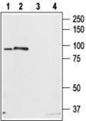
- Experimental details
- Western blot analysis of rat brain (lanes 1,3) and RBL cell (lanes 2,4) lysates: 1. Anti-STIM2 antibody, (1:200). 2. Anti-STIM2 antibody, preincubated with the control peptide antigen.
- Validation comment
- WB

Supportive validation
- Submitted by
- OriGene (provider)
- Main image
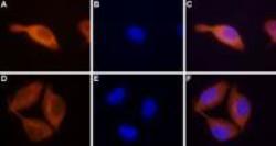
- Experimental details
- Expression of STIM2 in RBL cells. Immunocytochemical staining of paraformaldehyde-fixed and permeabilized rat basophilic leukemia (RBL) cells. A, D. Cells stained with Anti-STIM2 antibody, (1:100) followed by goat anti-rabbit-AlexaFluor-555 secondary antibody. B, E. Nuclear staining of cells using the cell-permeable dye Hoechst 33342. C. Merged image of panels A and B F. Merged image of panels D and E.
- Validation comment
- IF
Supportive validation
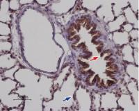
- Submitted by
- OriGene (provider)
- Main image
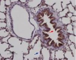
- Experimental details
- Expression of STIM2 in rat lung. Immunohistochemical staining of paraffin embedded rat lung sections using Anti-STIM2 antibody, (1:100). Staining is present in the respiratory epithelium of the bronchiole (red) as well as in the pneumonocytes of the alveolar wall (blue). Color reaction was obtained with SuperPicture HRP-conjugated polymer (Zymed) followed by DAB. Hematoxilin is used as the counterstain.
- Validation comment
- IHC