Explore
 Validate
 Learn
Learn Western blot
Antibody data
- Antibody Data
- Antigen structure
- References [0]
- Comments [0]
- Validations
- Western blot [3]
- Immunocytochemistry [1]
Submit
Validation data
Reference
Comment
Report error
- Product number
- 702729 - Provider product page

- Provider
- Invitrogen Antibodies
- Product name
- Aldolase A Recombinant Rabbit Monoclonal Antibody (21H7L6)
- Antibody type
- Monoclonal
- Antigen
- Synthetic peptide
- Description
- This antibody is predicted to react with Monkey, Cat, Rabbit
- Antibody clone number
- 21H7L6
- Concentration
- 0.5 mg/mL
No comments: Submit comment
Supportive validation
- Submitted by
- Invitrogen Antibodies (provider)
- Main image
- Experimental details
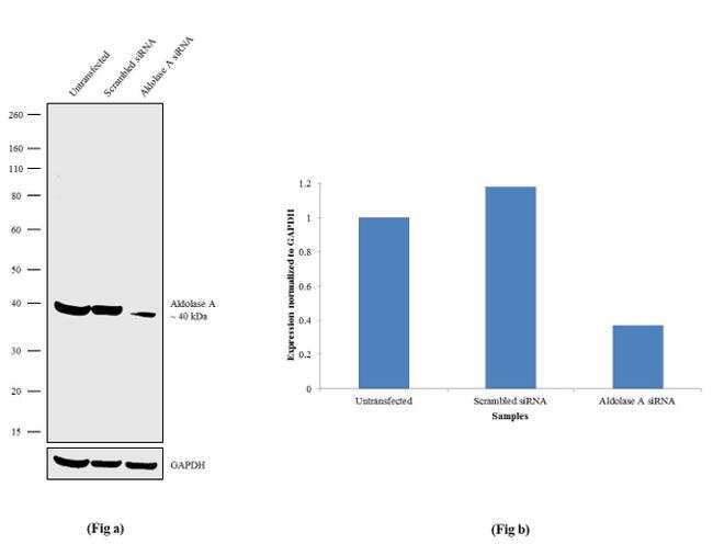
- Knockdown of Aldolase A was achieved by transfecting A549 cells with Aldolase A specific siRNA (Silencer® select Product # s72 + s71). Western blot analysis (Fig a) was performed using Whole cell extracts from the Aldolase A knockdown cells (Lane 3), non-specific scrambled siRNA transfected cells (Lane 2) and untransfected cells (Lane 1). The blots were probed with Anti-Aldolase A Recombinant Rabbit Monoclonal Antibody (Product # 702729, 1-3 µg/mL) and Goat anti-Rabbit IgG (H+L) Superclonal™ Secondary Antibody, HRP conjugate (Product # A27036, 0.25 µg/mL, 1:4000 dilution). Densitometric analysis of this Western blot is shown in histogram (Fig b). Loss of signal upon siRNA mediated knock down confirms that antibody is specific to Aldolase A.
- Submitted by
- Invitrogen Antibodies (provider)
- Main image

- Experimental details
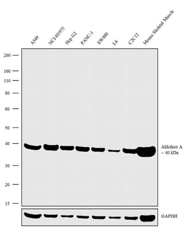
- Western blot analysis was performed on Whole cell extracts (30 µg lysate) of A549 (Lane 1), NCI-H1975 (Lane 2), Hep G2 (Lane 3), PANC-1 (Lane 4), SW480 (Lane 5), L6 (Lane 6), C2C12 (Lane 7) and tissue extracts of Mouse Skeletal Muscle (Lane 8). The blots were probed with Anti-Aldolase A Recombinant Rabbit Monoclonal Antibody (Product # 702729, 2.5 µg/mL) and detected by chemiluminescence using Goat anti-Rabbit IgG (H+L) Superclonal™ Secondary Antibody, HRP conjugate (Product # A27036, 0.25 µg/mL, 1:4000 dilution). A 40 kDa band corresponding to Aldolase A was observed across the cell lines and tissues tested. Known quantity of protein samples were electrophoresed using Novex®NuPAGE®4-12% Bis-Tris gel (Product # NP0322BOX), XCell SureLock™ Electrophoresis System (Product # EI0002) and Novex® Sharp Pre-Stained Protein Standard (Product # LC5800). Resolved proteins were then transferred onto a nitrocellulose membrane with iBlot® Dry Blotting System (Product # IB21001). The membrane was probed with the relevant primary and secondary Antibody following blocking with 5% skimmed milk. Chemiluminescent detection was performed using Pierce™ ECL Western blotting Substrate (Product # 32106).
- Submitted by
- Invitrogen Antibodies (provider)
- Main image

- Experimental details
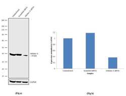
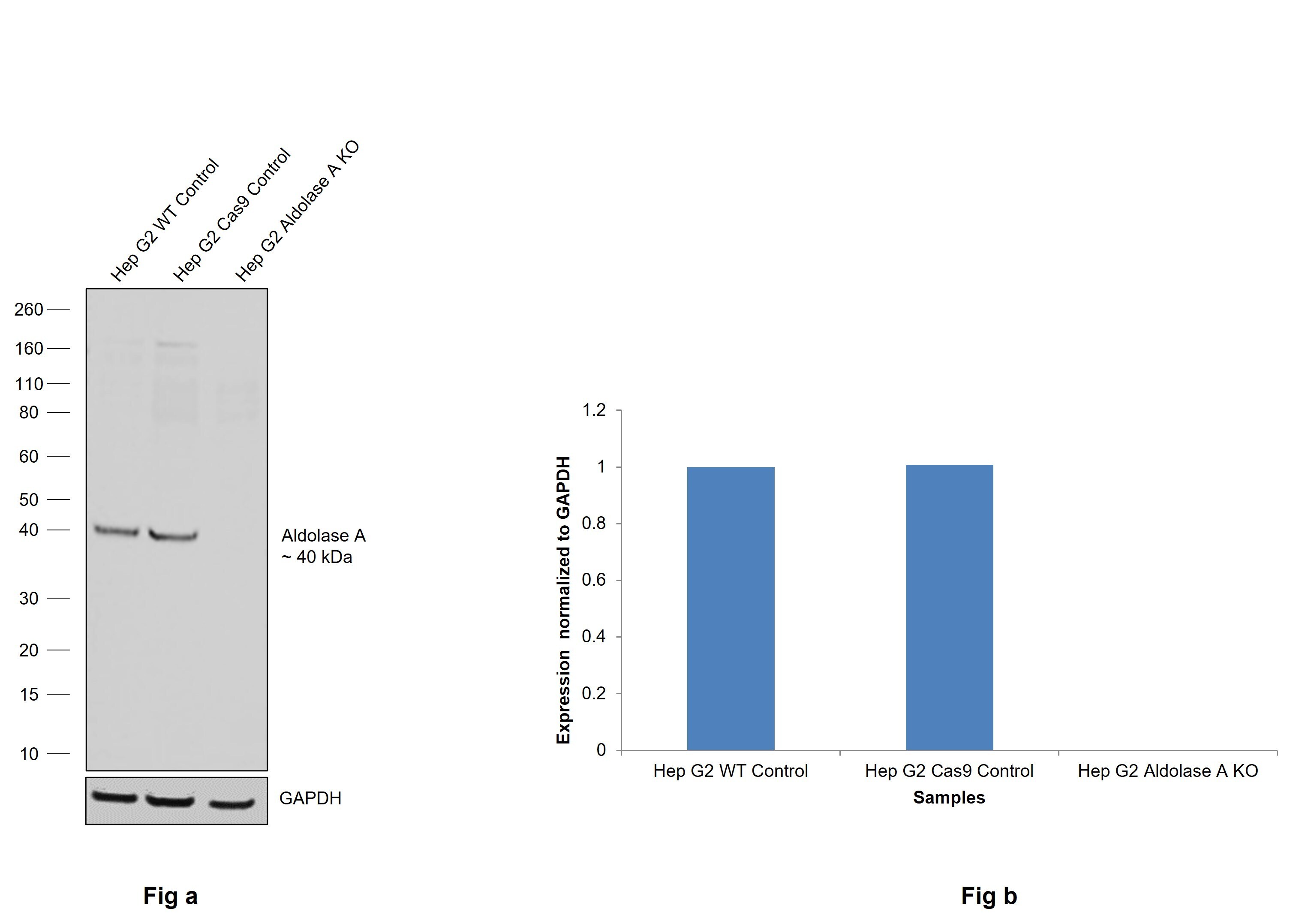
- Knockout of Aldolase A was achieved by CRISPR-Cas9 genome editing using LentiArray™ Lentiviral sgRNA (Product # A32042, Assay ID CRISPR891371_LV) and LentiArray Cas9 Lentivirus (Product # A32064). Western blot analysis of Aldolase A was performed by loading 30 µg of Hep G2 wild type (Lane 1), Hep G2 Cas9 (Lane 2) andHep G2 Aldolase A KO (Lane 3) whole cell extracts. The samples were electrophoresed using NuPAGE™ Novex™ 4-12% Bis-Tris Protein Gel (Product # NP0321BOX). Resolved proteins were then transferred onto a nitrocellulose membrane (Product # IB23001) by iBlot® 2 Dry Blotting System (Product # IB21001). The blot was probed with Anti-Aldolase A Recombinant Rabbit Monoclonal Antibody (21H7L6) (Product # 702729, 2.5 µg/mL dilution) and Goat anti-Rabbit IgG (H+L) Superclonal™ Recombinant Secondary Antibody, HRP (Product # A27036, 1:6000 dilution) using the iBright™ FL 1500 (Product # A44115). Chemiluminescent detection was performed using Novex® ECL Chemiluminescent Substrate Reagent Kit (Product # WP20005). Loss of signal upon CRISPR mediated knockout (KO) using the LentiArray™ CRISPR product line confirms that antibody is specific to Aldolase A.
Supportive validation
- Submitted by
- Invitrogen Antibodies (provider)
- Main image
- Experimental details
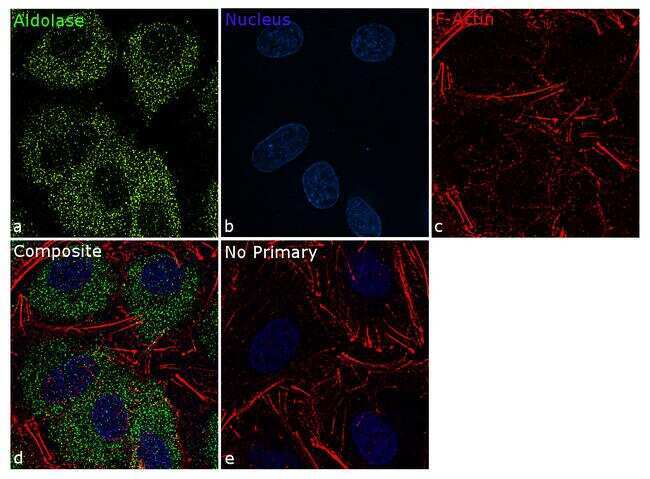
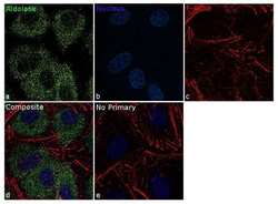
- For immunofluorescence analysis, A549 cells were fixed and permeabilized for detection of endogenous Aldolase using Anti- Aldolase Recombinant Rabbit Monoclonal Antibody (Product # 702729, 5 µg/mL) and labeled with Goat anti-Rabbit IgG (H+L) Superclonal™ Secondary Antibody, Alexa Fluor® 488 conjugate (Product # A27034, 1:2000). Panel a) shows representative cells that were stained for detection and localization of Aldolase protein (green), Panel b) is stained for nuclei (blue) using SlowFade® Gold Antifade Mountant with DAPI (Product # S36938). Panel c) represents cytoskeletal F-actin staining using Rhodamine Phalloidin (Product # R415, 1:300). Panel d) is a composite image of Panels a, b and c clearly demonstrating cytoplasmic localization of Aldolase A. Panel e) represents control cells with no primary antibody to assess background. The images were captured at 60X magnification.