Explore
 Validate
 Learn
Learn Western blot
Antibody data
- Antibody Data
- Antigen structure
- References [0]
- Comments [0]
- Validations
- Western blot [2]
- Immunocytochemistry [1]
- Immunohistochemistry [3]
Submit
Validation data
Reference
Comment
Report error
- Product number
- PA5-51951 - Provider product page

- Provider
- Invitrogen Antibodies
- Product name
- Cathepsin H Polyclonal Antibody
- Antibody type
- Polyclonal
- Antigen
- Recombinant full-length protein
- Description
- Immunogen sequence: LPSQAFEYIL YNKGIMGEDT YPYQGKDGYC KFQPGKAIGF VKDVANITIY DEEAMVEAVA LYNPVSFAFE VTQDFMMYRT GIYSSTSCHK TPDKVNHAVL AVGYGEKNGI PYWIVKNSWG PQWGMNGYFL
- Concentration
- 0.1 mg/mL
No comments: Submit comment
Supportive validation
- Submitted by
- Invitrogen Antibodies (provider)
- Main image

- Experimental details
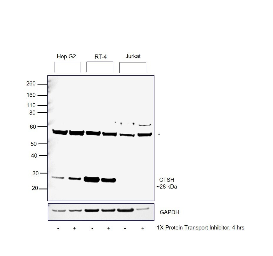
- Western blot was performed using Anti-Cathepsin H Polyclonal Antibody (Product # PA5-51951) and a 28 kDa band corresponding to mature form of Cathepsin H was observed across cell lines tested except Jurkat. Whole cell extracts (30 µg lysate) of Hep G2 (Lane 1), Hep G2 treated with 1X-PTI (Lane 2), RT-4 (Lane 3), RT-4 treated with 1X-PTI (Lane 4), Jurkat (Lane 5) and Jurkat treated with 1X-PTI (Lane 6) were electrophoresed using NuPAGE™ 4-12% Bis-Tris Protein Gel (Product # NP0322BOX). Resolved proteins were then transferred onto a nitrocellulose membrane (Product # IB23001) by iBlot® 2 Dry Blotting System (Product # IB21001). The blot was probed with the primary antibody (1:500 dilution) and detected by chemiluminescence with Goat anti-Rabbit IgG (H+L) Superclonal™ Recombinant Secondary Antibody, HRP (Product # A27036,1:20,000) using the iBright™ FL1500 Imaging System (Product # A44115). Chemiluminescent detection was performed using SuperSignal™ West Pico PLUS Chemiluminescent Substrate (Product # 34580). Hep G2 and RT-4 are high expressing cell models for CTSH, whereas, Jurkat does not express CTSH protein. An uncharacterized band (*) was observed around ~55 kDa across all cell lines.
- Submitted by
- Invitrogen Antibodies (provider)
- Main image

- Experimental details
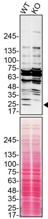
- Western blot of Pro-cathepsin H was performed by loading 100 µg of WT (lane 1) and CTSH CRISPR KO (lane 2) HEK293T cell lysates in RIPA buffer onto a 4-15% gradient polyacrylamide gel. Proteins on the blots were visualized with Ponceau staining (below immunoblot). Proteins were transferred to nitrocellulose membrane and blocked in 5% milk for 1 hr. CTSH was detected (designated by the black arrow) using a CTSH polyclonal antibody (Product # PA5-51951) at a dilution of 1:500 in 5% BSA in TBS with 0.1% Tween 20 (TBST) overnight at 4°C. The peroxidase-conjugated secondary antibody (Product # 65-6120) was diluted to 0.2 µg/mL in TBST with 5% milk for 1 hr. Chemiluminescent detection was performed using Pierce ECL Western Blotting Substrate (Product # 32106). Data courtesy of YCharOS Inc., an open science company with the mission of characterizing commercially available antibodies using knockout validation.
Supportive validation
- Submitted by
- Invitrogen Antibodies (provider)
- Main image

- Experimental details
- Immunofluorescent staining of Cathepsin H in human cell line U-2 OS using a Cathepsin H Polyclonal Antibody (Product # PA5-51951) shows localization to vesicles.

Supportive validation
- Submitted by
- Invitrogen Antibodies (provider)
- Main image

- Experimental details
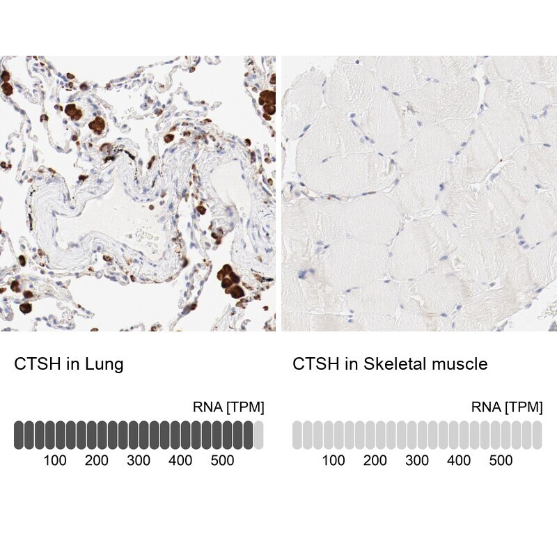
- Immunohistochemical staining of Cathepsin H in human lung and skeletal muscle tissues using Cathepsin H Polyclonal Antibody (Product # PA5-51951). Corresponding CTSH RNA-seq data are presented for the same tissues.
- Submitted by
- Invitrogen Antibodies (provider)
- Main image
- Experimental details
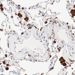
- Immunohistochemical staining of Cathepsin H in human lung using Cathepsin H Polyclonal Antibody (Product # PA5-51951) shows high expression.

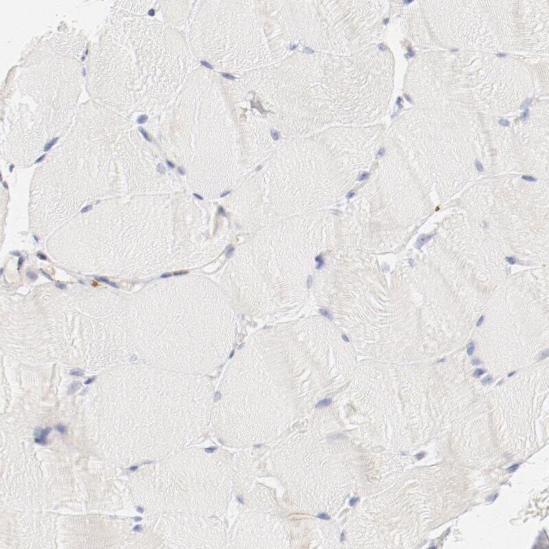
- Submitted by
- Invitrogen Antibodies (provider)
- Main image

- Experimental details
- Immunohistochemical staining of Cathepsin H in human skeletal muscle using Cathepsin H Polyclonal Antibody (Product # PA5-51951) shows low expression as expected.