Explore
 Validate
 Learn
Learn Western blot
Antibody data
- Antibody Data
- Antigen structure
- References [0]
- Comments [0]
- Validations
- Western blot [3]
- Immunocytochemistry [1]
- Immunohistochemistry [2]
- Other assay [1]
Submit
Validation data
Reference
Comment
Report error
- Product number
- MA5-27484 - Provider product page

- Provider
- Invitrogen Antibodies
- Product name
- IGF2BP3 Monoclonal Antibody (OTI6A3)
- Antibody type
- Monoclonal
- Antigen
- Recombinant full-length protein
- Reactivity
- Human, Mouse, Rat
- Host
- Mouse
- Isotype
- IgG
- Antibody clone number
- OTI6A3
- Vial size
- 100 µL
- Concentration
- 1 mg/mL
- Storage
- -20° C, Avoid Freeze/Thaw Cycles
No comments: Submit comment
Supportive validation
- Submitted by
- Invitrogen Antibodies (provider)
- Main image

- Experimental details
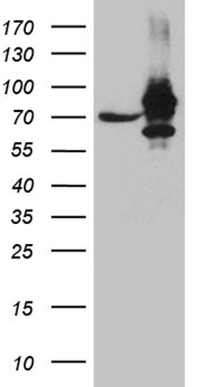
- Western blot analysis of IGF2BP3 in HEK293T cells in untransfected (Left lane) and transfected (Right lane) samples using 5 µg per lane. The samples were separated by SDS-PAGE and probed with IGF2BP3 (Product # MA5-27484) monoclonal antibody.
- Submitted by
- Invitrogen Antibodies (provider)
- Main image

- Experimental details
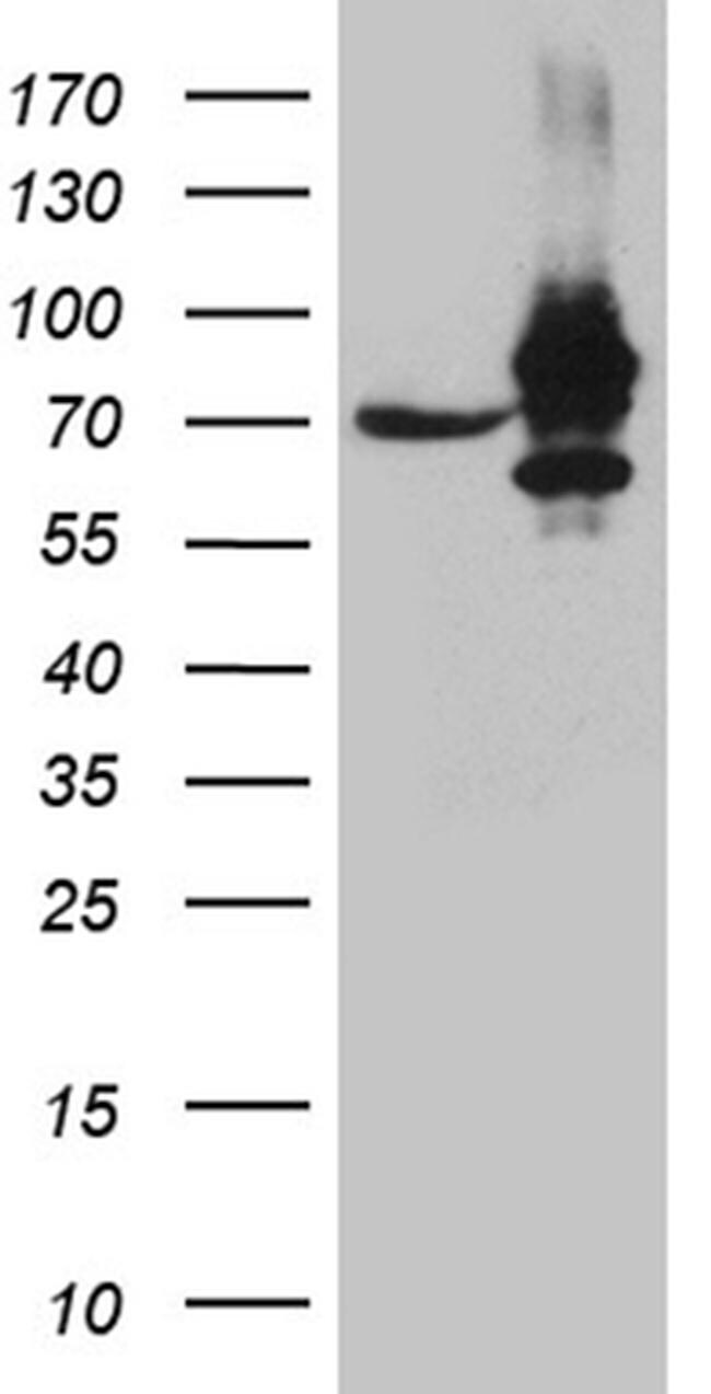
- Western blot analysis of IGF2BP3 in HEK293T cells in untransfected (Left lane) and transfected (Right lane) samples using 5 µg per lane. The samples were separated by SDS-PAGE and probed with IGF2BP3 (Product # MA5-27484) monoclonal antibody.
- Submitted by
- Invitrogen Antibodies (provider)
- Main image
- Experimental details
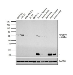
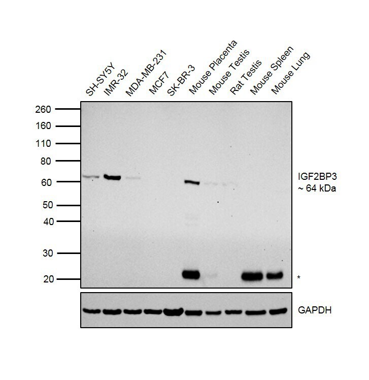
- Western blot was performed using Anti-IGF2BP3 Mouse Monoclonal Antibody (Product # MA5-27484) and a 64 kDa band corresponding to IGF2BP3 was observed in cell lines and tissues tested except MCF7, SK-BR-3, Mouse Lung and Mouse Spleen as reported. Whole cell extracts (30 µg lysate) of SH-SY5Y (Lane 1), IMR-32 (Lane 2), MDA-MB-231 (Lane 3), MCF7 (Lane 4), SK-BR-3 (Lane 5), Mouse Placenta (Lane 6), Mouse Testis (Lane 7), Rat Testis (Lane 8), Mouse Spleen (Lane 9) and Mouse Lung (Lane 10) were electrophoresed using Novex® NuPAGE® 4-12% Bis-Tris Protein Gel (Product # NP0322BOX). Resolved proteins were then transferred onto a nitrocellulose membrane (Product # IB23001) by iBlot® 2 Dry Blotting System (Product # IB21001). The blot was probed with the primary antibody (1:1000 dilution) and detected by chemiluminescence with Goat anti-Mouse IgG (H+L) Superclonal™ Recombinant Secondary Antibody, HRP (Product # A28177, 1:4000 dilution) using the iBright FL 1000 (Product # A32752). Chemiluminescent detection was performed using Novex® ECL Chemiluminescent Substrate Reagent Kit (Product # WP20005). A 25 kDa band (*) corresponding to circulating tissue IgG, was observed in mouse tissues.
Supportive validation
- Submitted by
- Invitrogen Antibodies (provider)
- Main image
- Experimental details
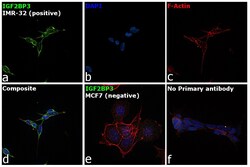
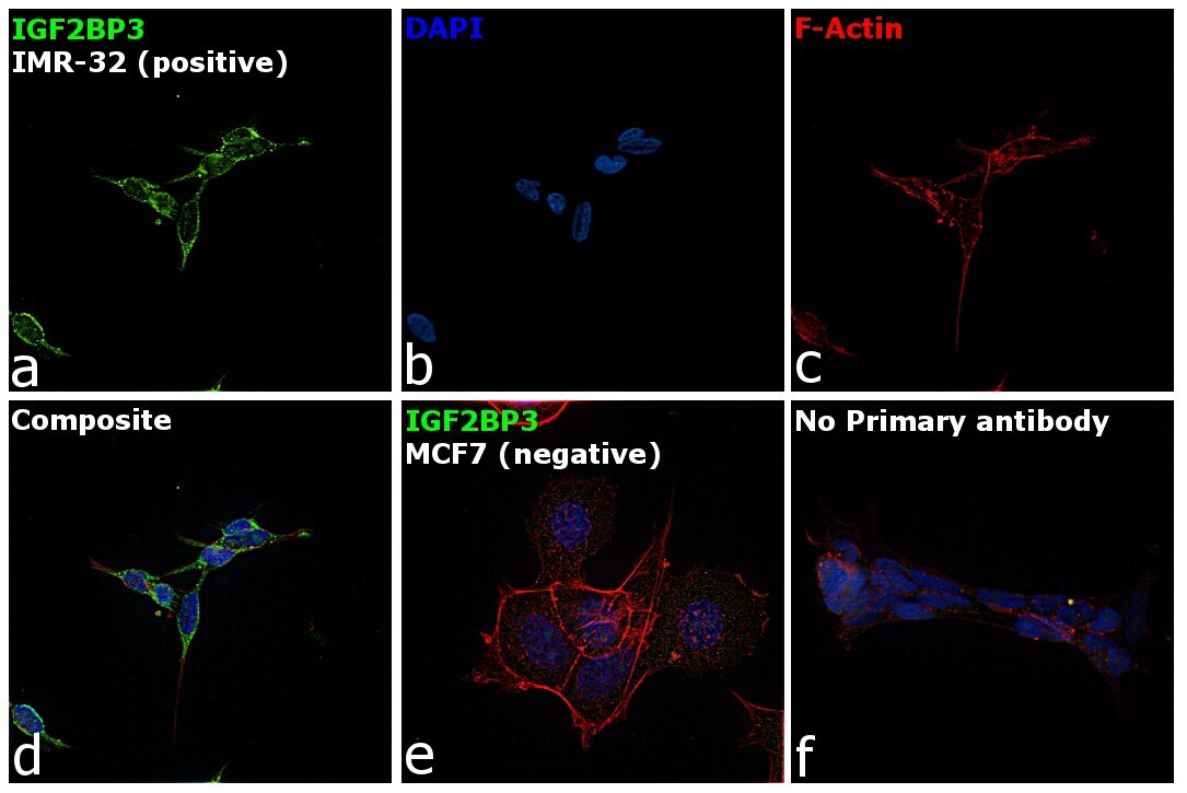
- Immunofluorescence analysis of IGF2BP3 was performed using IMR-32 and MCF7 cells. The cells were fixed with 4% paraformaldehyde for 10 minutes, permeabilized with 0.1% Triton™ X-100 for 15 minutes, and blocked with 2% BSA for 1 hour at room temperature. The cells were labeled with IGF2BP3 Mouse Monoclonal Antibody (Product # MA5-27484) at 1:100 dilution in 0.1% BSA and incubated overnight at 4 degree and then labeled with Goat anti-Mouse IgG (H+L) Highly Cross-Adsorbed Secondary Antibody, Alexa Fluor Plus 488 (Product # A32723) at a dilution of 1:2000 for 45 minutes at room temperature (Panel a: green) in IMR-32 cells. Nuclei (Panel b: blue) were stained with ProLong™ Diamond Antifade Mountant with DAPI (Product # P36962). F-actin (Panel c: red) was stained with Rhodamine Phalloidin (Product # R415, 1:300). Panel d represents the merged image of IMR-32 cells, which is a positive model for IGF2BP3 expression showing a cytoplasmic and nuclear localization. Panel e represents the merged image ofMCF7 cells, that are null for IGF2BP3 protein expression. Panel f represents control cells with no primary antibody to assess background. The images were captured at 60X magnification.
Supportive validation
- Submitted by
- Invitrogen Antibodies (provider)
- Main image
- Experimental details
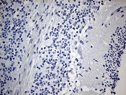
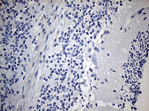
- Immunohistochemistry was performed on paraffin-embedded human embryonic cerebellum tissue. To expose target proteins, heat-induced epitope retrieval by 1mM EDTA in 10mM Tris buffer (pH8.5) at 120°C for 3 min. Following antigen retrieval, tissues were probed with a IGF2BP3 monoclonal antibody (Product # MA5-27484) at a dilution of 1:150.
- Submitted by
- Invitrogen Antibodies (provider)
- Main image
- Experimental details
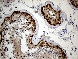
- Immunohistochemistry was performed on paraffin-embedded human testicle tissue. To expose target proteins, heat-induced epitope retrieval by 1mM EDTA in 10mM Tris buffer (pH8.5) at 120°C for 3 min. Following antigen retrieval, tissues were probed with a IGF2BP3 monoclonal antibody (Product # MA5-27484) at a dilution of 1:150.

Supportive validation
- Submitted by
- Invitrogen Antibodies (provider)
- Main image

- Experimental details
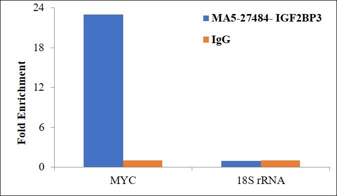
- RNA Immunoprecipitation (RIP) assay of endogenous IGF2BP3 protein using Anti-IGF2BP3 Antibody: RIP assay was performed using Anti-IGF2BP3 Monoclonal Antibody (Product # MA5-27484) 5 µg, on whole cell lysate from Hep G2 cells. Normal Mouse IgG was used as a negative IP control. RNA purified by RiboPure™ RNA Purification Kit (Product # AM1924) was analyzed by RT-PCR using the Power SYBR® Green RNA-to-CT™ 1-Step Kit (Product # 4389986) with the primers pairs over MYC exon1 and 18S rRNA. Data is presented as fold enrichment of the antibody signal versus the negative control IgG using the comparative CT method.