 Explore
 Validate
 Learn
Learn Western blot
Antibody data
- Antibody Data
- Antigen structure
- References [1]
- Comments [0]
- Validations
- Western blot [1]
- Immunocytochemistry [1]
- Chromatin Immunoprecipitation [1]
Submit
Validation data
Reference
Comment
Report error
- Product number
- PA5-27472 - Provider product page

- Provider
- Invitrogen Antibodies
- Product name
- CHD4 Polyclonal Antibody
- Antibody type
- Polyclonal
- Antigen
- Recombinant protein fragment
- Description
- Recommended positive controls: Jurkat, Raji, K562.
- Concentration
- 0.84 mg/mL
Submitted references NuRD subunit CHD4 regulates super-enhancer accessibility in rhabdomyosarcoma and represents a general tumor dependency.
Marques JG, Gryder BE, Pavlovic B, Chung Y, Ngo QA, Frommelt F, Gstaiger M, Song Y, Benischke K, Laubscher D, Wachtel M, Khan J, Schäfer BW
eLife 2020 Aug 3;9
eLife 2020 Aug 3;9
No comments: Submit comment
Supportive validation
- Submitted by
- Invitrogen Antibodies (provider)
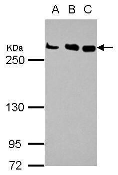
- Main image

- Experimental details
- Western Blot using CHD4 Polyclonal Antibody (Product # PA5-27472). Sample (30 µg of whole cell lysate). Lane A: Jurkat. Lane B: Raji. Lane C: K562 . 5% SDS PAGE. CHD4 Polyclonal Antibody (Product # PA5-27472) diluted at 1:1,000.
Supportive validation
- Submitted by
- Invitrogen Antibodies (provider)
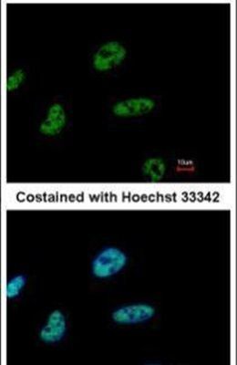
- Main image
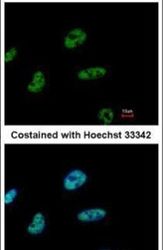
- Experimental details
- Immunofluorescent analysis of CHD4 in paraformaldehyde-fixed HeLa cells using a CHD4 polyclonal antibody (Product # PA5-27472) at a 1:500 dilution.
Supportive validation
- Submitted by
- Invitrogen Antibodies (provider)
- Main image

- Experimental details
- Chromatin Immunoprecipitation (ChIP) assay of endogenous CHD4 protein using Anti-CHD4 Antibody: ChIP was performed using Anti-CHD4 Rabbit Polyclonal Antibody (Product # PA5-27472, 5 µg) on sheared chromatin from HeLa cells using the MAGnify ChIP System kit (Product # 49-2024). Normal Rabbit IgG was used as a negative IP control. The purified DNA was analyzed by qPCR using primers binding to PKN3 and GAPDH transcriptional start site, CDKN1A exon 2 and SAT2 satellite repeats. Data is presented as fold enrichment of the antibody signal versus the negative control IgG using the comparative CT method.
