Explore
 Validate
 Learn
Learn37-2700
antibody from Invitrogen Antibodies
Targeting: CSPG4
CSPG4A, HMW-MAA, MCSP, MCSPG, MEL-CSPG, MSK16, NG2
 ELISA
ELISA Other assay
Antibody data
- Antibody Data
- Antigen structure
- References [9]
- Comments [0]
- Validations
- Other assay [9]
Submit
Validation data
Reference
Comment
Report error
- Product number
- 37-2700 - Provider product page

- Provider
- Invitrogen Antibodies
- Product name
- NG2 Monoclonal Antibody (D120.43/D4.11/N143.8/N109.6)
- Antibody type
- Monoclonal
- Antigen
- Purifed from natural sources
- Reactivity
- Human, Rat
- Host
- Mouse
- Isotype
- IgG
- Antibody clone number
- D120.43/D4.11/N143.8/N109.6
- Vial size
- 100 µg
- Concentration
- 0.5 mg/mL
- Storage
- -20°C
Submitted references TUBB4A mutations result in both glial and neuronal degeneration in an H-ABC leukodystrophy mouse model.
Resistance to retinopathy development in obese, diabetic and hypertensive ZSF1 rats: an exciting model to identify protective genes.
CD200 restrains macrophage attack on oligodendrocyte precursors via toll-like receptor 4 downregulation.
Phenotype overlap in glial cell populations: astroglia, oligodendroglia and NG-2(+) cells.
Spinal cord injury induces a long-lasting upregulation of interleukin-1β in astrocytes around the central canal.
Perivascular-derived stem cells with neural crest characteristics are involved in tendon repair.
Spatio-temporal expression pattern of frizzled receptors after contusive spinal cord injury in adult rats.
Chemokine expression in the white matter spinal cord precursor niche after force-defined spinal cord contusion injuries in adult rats.
Subcellular localization of transporters along the rat blood-brain barrier and blood-cerebral-spinal fluid barrier by in vivo biotinylation.
Sase S, Almad AA, Boecker CA, Guedes-Dias P, Li JJ, Takanohashi A, Patel A, McCaffrey T, Patel H, Sirdeshpande D, Curiel J, Shih-Hwa Liu J, Padiath Q, Holzbaur EL, Scherer SS, Vanderver A
eLife 2020 May 28;9
eLife 2020 May 28;9
Resistance to retinopathy development in obese, diabetic and hypertensive ZSF1 rats: an exciting model to identify protective genes.
Caolo V, Roblain Q, Lecomte J, Carai P, Peters L, Cuijpers I, Robinson EL, Derks K, Sergeys J, Noël A, Jones EAV, Moons L, Heymans S
Scientific reports 2018 Aug 9;8(1):11922
Scientific reports 2018 Aug 9;8(1):11922
CD200 restrains macrophage attack on oligodendrocyte precursors via toll-like receptor 4 downregulation.
Hayakawa K, Pham LD, Seo JH, Miyamoto N, Maki T, Terasaki Y, Sakadžić S, Boas D, van Leyen K, Waeber C, Kim KW, Arai K, Lo EH
Journal of cerebral blood flow and metabolism : official journal of the International Society of Cerebral Blood Flow and Metabolism 2016 Apr;36(4):781-93
Journal of cerebral blood flow and metabolism : official journal of the International Society of Cerebral Blood Flow and Metabolism 2016 Apr;36(4):781-93
Phenotype overlap in glial cell populations: astroglia, oligodendroglia and NG-2(+) cells.
Alghamdi B, Fern R
Frontiers in neuroanatomy 2015;9:49
Frontiers in neuroanatomy 2015;9:49
Spinal cord injury induces a long-lasting upregulation of interleukin-1β in astrocytes around the central canal.
Paniagua-Torija B, Arevalo-Martin A, Molina-Holgado E, Molina-Holgado F, Garcia-Ovejero D
Neuroscience 2015 Jan 22;284:283-289
Neuroscience 2015 Jan 22;284:283-289
Perivascular-derived stem cells with neural crest characteristics are involved in tendon repair.
Xu W, Sun Y, Zhang J, Xu K, Pan L, He L, Song Y, Njunge L, Xu Z, Chiang MY, Sung KL, Chuong CM, Yang L
Stem cells and development 2015 Apr 1;24(7):857-68
Stem cells and development 2015 Apr 1;24(7):857-68
Spatio-temporal expression pattern of frizzled receptors after contusive spinal cord injury in adult rats.
Gonzalez P, Fernandez-Martos CM, Gonzalez-Fernandez C, Arenas E, Rodriguez FJ
PloS one 2012;7(12):e50793
PloS one 2012;7(12):e50793
Chemokine expression in the white matter spinal cord precursor niche after force-defined spinal cord contusion injuries in adult rats.
Knerlich-Lukoschus F, von der Ropp-Brenner B, Lucius R, Mehdorn HM, Held-Feindt J
Glia 2010 Jun;58(8):916-31
Glia 2010 Jun;58(8):916-31
Subcellular localization of transporters along the rat blood-brain barrier and blood-cerebral-spinal fluid barrier by in vivo biotinylation.
Roberts LM, Black DS, Raman C, Woodford K, Zhou M, Haggerty JE, Yan AT, Cwirla SE, Grindstaff KK
Neuroscience 2008 Aug 13;155(2):423-38
Neuroscience 2008 Aug 13;155(2):423-38
No comments: Submit comment
Supportive validation
- Submitted by
- Invitrogen Antibodies (provider)
- Main image
- Experimental details
- NULL

- Submitted by
- Invitrogen Antibodies (provider)
- Main image
- Experimental details
- NULL
- Submitted by
- Invitrogen Antibodies (provider)
- Main image
- Experimental details
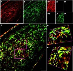
- Figure 1 GFAP and NG-2 co-localization in P10 RON glial cells. (A) GFAP immuno-reactivity (red). (B) NG-2 immuno-reactivity (green). (C) Overlay with boxed areas shown at higher gain as 3-D projections in (C1) and (C2) . Note the NG-2 co-localization (red or orange regions e.g., light blue arrows) in parts of cells that may also have regions that are only GFAP(+) (green regions e.g., dark blue arrow). (D) Controls showing GFAP staining (D1) and absence of NG-2 staining (D2) when the NG-2 antibody was omitted from the otherwise identical protocol; NG-2 staining (D3) and no GFAP staining (D4) when the GFAP antibody was omitted, and the absence of any staining when both primary antibodies were omitted ( D5,D6 ). All images were collected and displayed using identical settings. Bar = 10 mum.

- Submitted by
- Invitrogen Antibodies (provider)
- Main image
- Experimental details
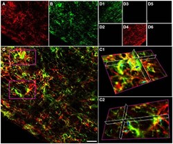
- Figure 2 GFAP and NG-2 co-localization in the adult RON. (A) GFAP immuno-reactivity (red). (B) NG-2 immuno-reactivity (green). (C) Overlay with boxed areas shown at higher gain as 3-D projections in (C1) and (C2) . Note the NG-2 co-localization (red or orange, e.g., light blue arrows) in parts of cells that may express GFAP alone in other regions, and in cells that are only GFAP(+) (e.g., dark blue arrow). (D) Controls showing NG-2 staining (D1) and absence of GFAP staining (D2) when the GFAP antibody was omitted from the otherwise identical protocol; GFAP staining (D4) and no NG-2 staining (D3) when the NG-2 antibody was omitted, and the absence of any staining when both primary antibodies were omitted ( D5,D6 ). All images were collected and displayed using identical settings. Bar = 10 mum.
- Submitted by
- Invitrogen Antibodies (provider)
- Main image
- Experimental details
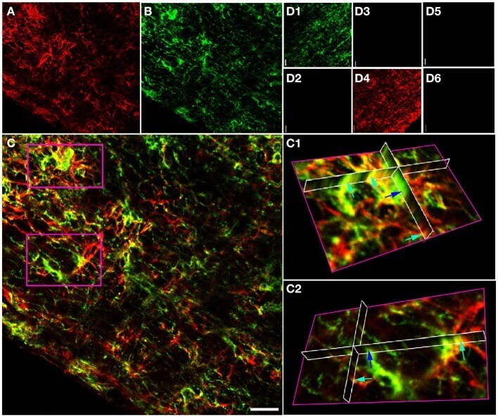
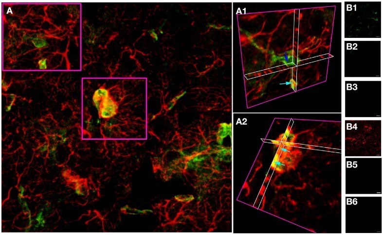
- Figure 3 GFAP and NG-2 co-localization in adult cortical gray matter. (A) GFAP immuno-reactivity (red) and NG-2 immuno-reactivity (green), with the two boxed areas shown at higher gain as 3-D projections in (A1) and (A2) . Note the NG-2 co-localization (e.g., light blue arrows) in parts of cells that may also have regions that are only GFAP(+), while other NG-2(+) cells are GFAP(-) (e.g., dark blue arrow). (B) Controls showing GFAP staining (B1) and absence of NG-2 staining (B2) when the NG-2 antibody was omitted from the otherwise identical protocol; NG-2 staining (B4) and no GFAP staining (B3) when the GFAP antibody was omitted, and the absence of any staining when both primary antibodies were omitted ( B5,B6 ). All images were collected and displayed using identical settings. Bar = 10 mum.
- Submitted by
- Invitrogen Antibodies (provider)
- Main image
- Experimental details
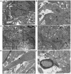
- Figure 4 NG-2 immuno-gold labeling in P10 RON. (A,B) Two closely apposed glial soma (""1"" and ""2""). Cell ""1"" has features typical of an early cell of the oligodendroglial lineage including an ovoid nucleus and narrow bore ER. Cell ""2"" has features that are typical of astrocytes in this preparation. The boxed area is shown at higher gain in ( B ). Note the gold particles (some indicated by arrows) which identify this cell as NG-2(+). A lobular nuclear morphology with clustered chromatin under the nuclear envelope and a wide bore ER (arrow heads) are astrocyte features. The cytoplasm also contains microtubules (e.g., asterisk). Glial filaments cannot be positively identified in this cell. (C,D) Another NG-2(+) cell with astrocyte features which does express glial filaments (double arrows). Boxed area shown at higher gain in (D) . (E) High-gain micrograph of NG-2 staining in glial processes (arrows) which contains glia filaments (arrowhead). (F) An example of NG-2(+) (arrows) oligodendrocyte processes ensheathing an axon.

- Submitted by
- Invitrogen Antibodies (provider)
- Main image

- Experimental details
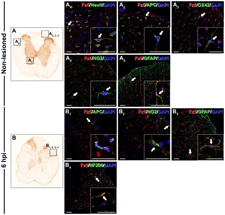
- Figure 9 Cellular protein expression pattern of Frizzled 5 in non-lesioned spinal cords and at 6 hpi. This figure shows representative images obtained from the microscopic evaluation of sections processed by double immunohistochemistry to visualise Frizzled (Fz) 5 in astrocytes (glial fibrillary acidic protein (GFAP)), neurons (neuronal nuclei (NeuN)), oligodendrocytes (adenomatous polyposis coli (APC)), axons (neurofilament 200 (NF200)), microglia/macrophages (OX-42) and glial progenitors (NG2) in the non-lesioned spinal cords (9A, A 1 , A 2 , A 3 , A 4 and A 5 ) and at 6 hours post-injury (hpi) (9B, B 1 , B 2 , B 3 and B 4 ). The squares in the images showing the entire spinal cord sections (9A and B) correspond to the areas of higher magnification. Scale bars = 40 um.
- Submitted by
- Invitrogen Antibodies (provider)
- Main image
- Experimental details
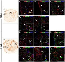
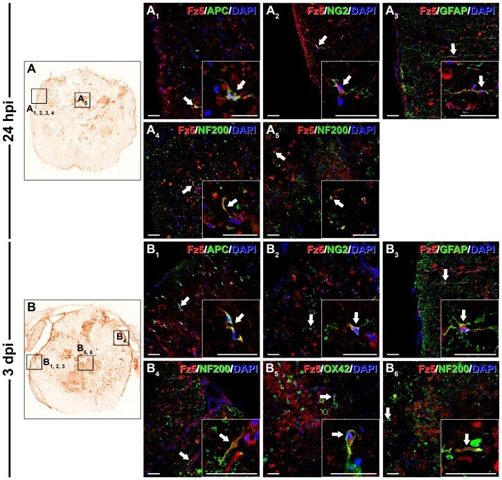
- Figure 10 Cellular protein expression pattern of Frizzled 5 at 24 hpi and 3 dpi. This figure shows representative images obtained from the microscopic evaluation of sections processed by double immunohistochemistry to visualise Frizzled (Fz) 5 in astrocytes (glial fibrillary acidic protein (GFAP)), neurons (neuronal nuclei (NeuN)), oligodendrocytes (adenomatous polyposis coli (APC)), axons (neurofilament 200 (NF200)), microglia/macrophages (OX-42) and glial progenitors (NG2) at 24 hours post-injury (hpi) (10A, A 1 , A 2 , A 3 , A 4 and A 5 ) and 3 days post-injury (dpi) (10B, B 1 , B 2 , B 3 , B 4 , B 5 and B 6 ). The squares in the images showing the entire spinal cord sections (10A and B) correspond to the areas of higher magnification. Scale bars = 40 um.
- Submitted by
- Invitrogen Antibodies (provider)
- Main image
- Experimental details
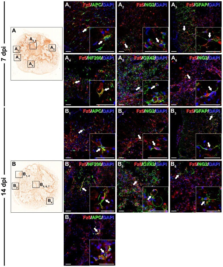
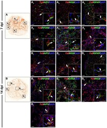
- Figure 11 Cellular protein expression pattern of Frizzled 5 at 7 and 14 dpi. This figure shows representative images obtained from the microscopic evaluation of sections processed by double immunohistochemistry to visualise Frizzled (Fz) 5 in astrocytes (glial fibrillary acidic protein (GFAP)), neurons (neuronal nuclei (NeuN)), oligodendrocytes (adenomatous polyposis coli (APC)), axons (neurofilament 200 (NF200)), microglia/macrophages (OX-42) and glial progenitors (NG2) at 7 (11A, A 1 , A 2 , A 3 , A 4 , A 5 and A 6 ) and 14 days post-injury (dpi) (11B, B 1 , B 2 , B 3 , B 4 , B 5 , B 6 and B 7 ). The squares in the images showing the entire spinal cord sections (11A and B) correspond to the areas of higher magnification. Scale bars = 40 um.