Explore
 Validate
 Learn
LearnPA5-30081
antibody from Invitrogen Antibodies
Targeting: LMAN1
ERGIC-53, ERGIC53, F5F8D, FMFD1, gp58, MCFD1, MR60
 Western blot
Antibody data
- Antibody Data
- Antigen structure
- References [0]
- Comments [0]
- Validations
- Western blot [3]
- Immunohistochemistry [2]
Submit
Validation data
Reference
Comment
Report error
- Product number
- PA5-30081 - Provider product page

- Provider
- Invitrogen Antibodies
- Product name
- LMAN1 Polyclonal Antibody
- Antibody type
- Polyclonal
- Antigen
- Recombinant protein fragment
- Description
- Recommended positive controls: HeLa, HepG2, HCT116.
- Concentration
- 0.82 mg/mL
No comments: Submit comment
Supportive validation
- Submitted by
- Invitrogen Antibodies (provider)
- Main image

- Experimental details
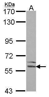
- Western Blot using LMAN1 Polyclonal Antibody (Product # PA5-30081). Sample (30 µg of whole cell lysate). Lane A: Hela . 7.5% SDS PAGE. LMAN1 Polyclonal Antibody (Product # PA5-30081) diluted at 1:1,000.
- Submitted by
- Invitrogen Antibodies (provider)
- Main image

- Experimental details
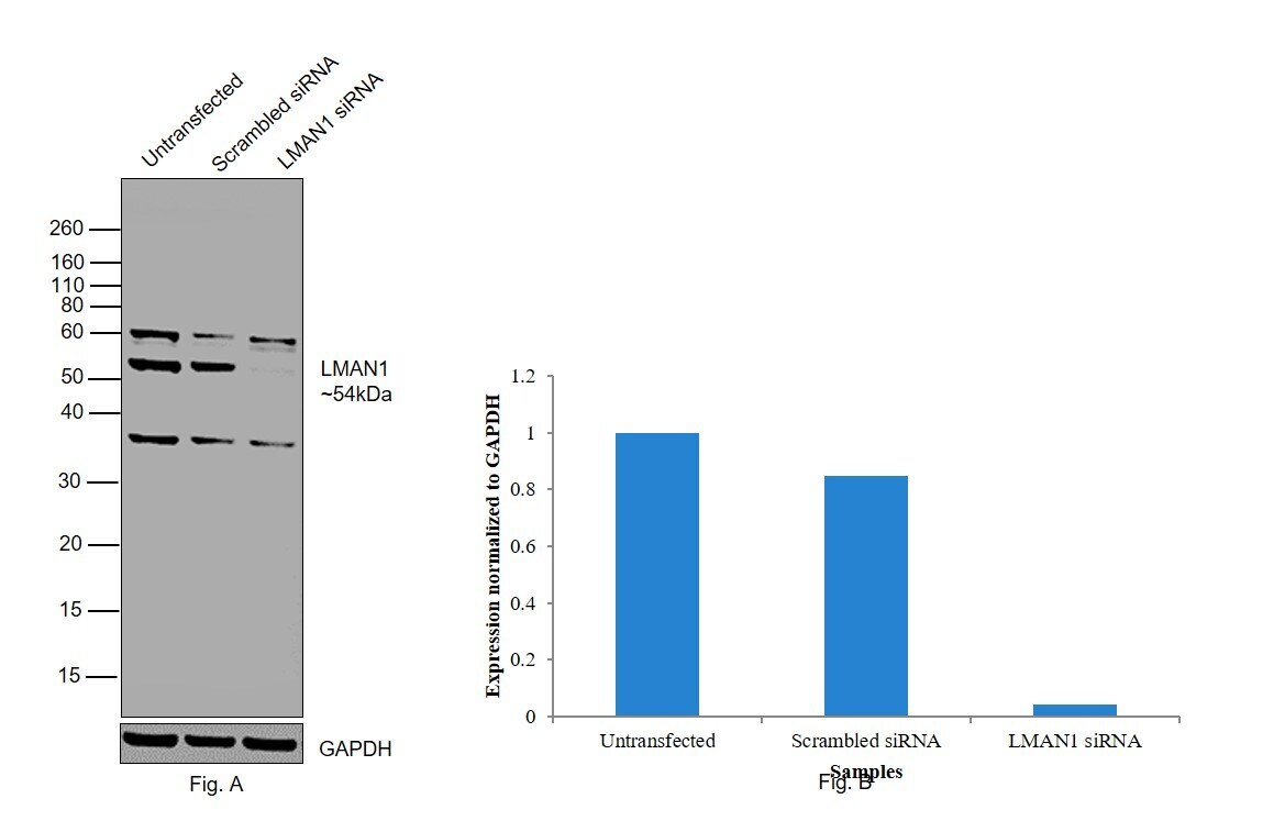
- Knockdown of LMAN1 was achieved by transfecting HeLa with LMAN1 specific siRNAs (Silencer® select Product # s8218, s8220). Western blot analysis (Fig. a) was performed using whole cell extracts from the LMAN1 knockdown cells (Lane 3), non-specific scrambled siRNA transfected cells (Lane 2) and untransfected cells (Lane 1). The blot was probed with LMAN1 Polyclonal Antibody (Product # PA5-30081, 1:1000 dilution) and Goat anti-Rabbit IgG (H+L) Superclonal™ Recombinant Secondary Antibody, HRP (Product # A27036, 1:4000 dilution). Densitometric analysis of this western blot is shown in histogram (Fig. b). Decrease in signal upon siRNA mediated knock down confirms that antibody is specific to LMAN1. Few uncharacterized bands were also observed at different molecular weights.
- Submitted by
- Invitrogen Antibodies (provider)
- Main image
- Experimental details
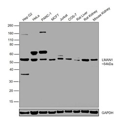
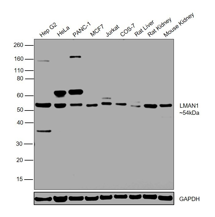
- Western blot was performed using Anti-LMAN1 Polyclonal Antibody (Product # PA5-30081) and a 54kDa band corresponding to LMAN1 was observed in all tested cell models and tissue lysates along with few uncharacterized bands. Whole cell extracts (30ug) of Hep G2 (Lane 1), HeLa (Lane 2), PANC-1 (Lane 3), MCF7 (Lane 4), Jurkat (Lane 5), COS-7 (Lane 6), tissue lysates of Rat Liver (Lane 7), Rat Kidney (Lane 8) and Mouse Kidney (Lane 9) were electrophoresed using Novex® NuPAGE® 4-12 % Bis-Tris gel (Product # NP0322BOX). Resolved proteins were then transferred onto a nitrocellulose membrane (Product # IB23001) by iBlot® 2 Dry Blotting System (Product # IB21001). The blot was probed with the primary antibody (1:1000 dilution) and detected by chemiluminescence with Goat anti-Rabbit IgG (H+L) Superclonal™ Recombinant Secondary Antibody, HRP (Product # A27036, 1:4000 dilution) using the iBright FL 1000 (Product # A32752). Chemiluminescent detection was performed using Novex® ECL Chemiluminescent Substrate Reagent Kit (Product # WP20005).
Supportive validation
- Submitted by
- Invitrogen Antibodies (provider)
- Main image
- Experimental details
- Immunohistochemical analysis of paraffin-embedded human breast cancer, using LMAN1 (Product # PA5-30081) antibody at 1:500 dilution. Antigen Retrieval: EDTA based buffer, pH 8.0, 15 min.

- Submitted by
- Invitrogen Antibodies (provider)
- Main image

- Experimental details
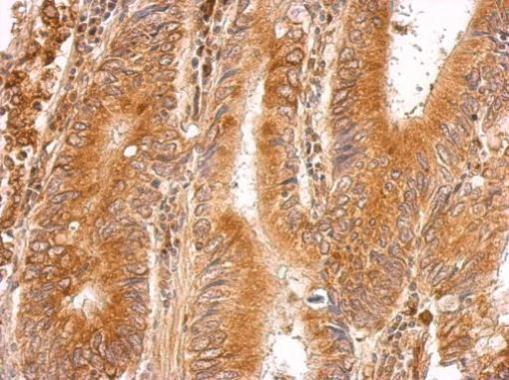
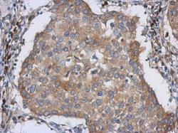
- LMAN1 Polyclonal Antibody detects LMAN1 protein at cytosol on human gastric cancer by immunohistochemical analysis. Sample: Paraffin-embedded gastric cancer. LMAN1 Polyclonal Antibody (Product # PA5-30081) dilution: 1:500. Antigen Retrieval: EDTA based buffer, pH 8.0, 15 min.