Explore
 Validate
 Learn
Learn45-8900
antibody from Invitrogen Antibodies
Targeting: AURKA
AIK, ARK1, AurA, BTAK, PPP1R47, STK15, STK6, STK7
 Western blot
 Immunoprecipitation
Antibody data
- Antibody Data
- Antigen structure
- References [6]
- Comments [0]
- Validations
- Western blot [3]
- Immunocytochemistry [1]
- Other assay [1]
Submit
Validation data
Reference
Comment
Report error
- Product number
- 45-8900 - Provider product page

- Provider
- Invitrogen Antibodies
- Product name
- Aurora A Monoclonal Antibody (35C1)
- Antibody type
- Monoclonal
- Antigen
- Recombinant full-length protein
- Reactivity
- Human, Mouse, Rat, Bovine, Canine, Porcine
- Host
- Mouse
- Isotype
- IgG
- Antibody clone number
- 35C1
- Vial size
- 100 µg
- Concentration
- 0.5 mg/mL
- Storage
- Store at 4°C short term. For long term storage, store at -20°C, avoiding freeze/thaw cycles.
Submitted references Myocardin-related transcription factor and serum response factor regulate cilium turnover by both transcriptional and local mechanisms.
Targeting Aurora A Kinase (AAK) in Platinum-Resistant High Grade Serous Ovarian Cancer.
A novel mitosis-specific Cep215 domain interacts with Cep192 and phosphorylated Aurora A for organization of spindle poles.
Quantitative Temporal Viromics of an Inducible HIV-1 Model Yields Insight to Global Host Targets and Phospho-Dynamics Associated with Protein Vpr.
14-3-3γ Prevents Centrosome Amplification and Neoplastic Progression.
Aurora-A mediated histone H3 phosphorylation of threonine 118 controls condensin I and cohesin occupancy in mitosis.
Speight P, Rozycki M, Venugopal S, Szászi K, Kofler M, Kapus A
iScience 2021 Jul 23;24(7):102739
iScience 2021 Jul 23;24(7):102739
Targeting Aurora A Kinase (AAK) in Platinum-Resistant High Grade Serous Ovarian Cancer.
Ganapathi RN, Norris EJ, Sutker AP, Klotz KE, Ganapathi MK
Frontiers in oncology 2020;10:1354
Frontiers in oncology 2020;10:1354
A novel mitosis-specific Cep215 domain interacts with Cep192 and phosphorylated Aurora A for organization of spindle poles.
Kuriyama R, Fisher CR
Journal of cell science 2020 Dec 29;133(24)
Journal of cell science 2020 Dec 29;133(24)
Quantitative Temporal Viromics of an Inducible HIV-1 Model Yields Insight to Global Host Targets and Phospho-Dynamics Associated with Protein Vpr.
Lapek JD Jr, Lewinski MK, Wozniak JM, Guatelli J, Gonzalez DJ
Molecular & cellular proteomics : MCP 2017 Aug;16(8):1447-1461
Molecular & cellular proteomics : MCP 2017 Aug;16(8):1447-1461
14-3-3γ Prevents Centrosome Amplification and Neoplastic Progression.
Mukhopadhyay A, Sehgal L, Bose A, Gulvady A, Senapati P, Thorat R, Basu S, Bhatt K, Hosing AS, Balyan R, Borde L, Kundu TK, Dalal SN
Scientific reports 2016 Jun 2;6:26580
Scientific reports 2016 Jun 2;6:26580
Aurora-A mediated histone H3 phosphorylation of threonine 118 controls condensin I and cohesin occupancy in mitosis.
Wike CL, Graves HK, Hawkins R, Gibson MD, Ferdinand MB, Zhang T, Chen Z, Hudson DF, Ottesen JJ, Poirier MG, Schumacher J, Tyler JK
eLife 2016 Feb 16;5:e11402
eLife 2016 Feb 16;5:e11402
No comments: Submit comment
Supportive validation
- Submitted by
- Invitrogen Antibodies (provider)
- Main image
- Experimental details
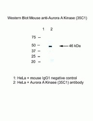
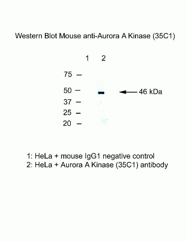
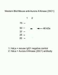
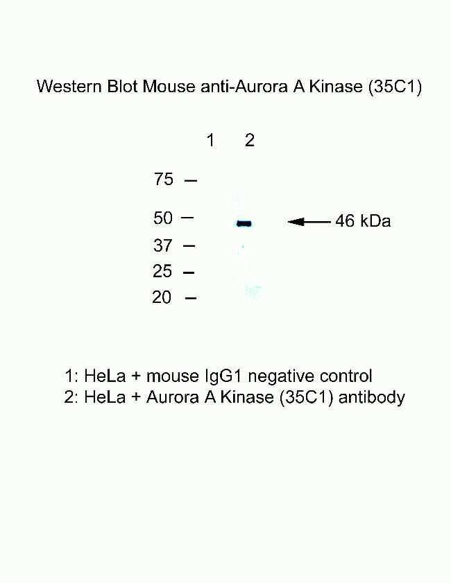
- Western blot analysis of Aurora A using a monoclonal antibody (Product # 45-8900).
- Submitted by
- Invitrogen Antibodies (provider)
- Main image
- Experimental details
- Western blot analysis of Aurora A using a monoclonal antibody (Product # 45-8900).
- Submitted by
- Invitrogen Antibodies (provider)
- Main image

- Experimental details
- Western blot analysis of Aurora A was performed by loading 30 µg of Jurkat (lane1), K562 (lane2) and MCF7 (lane3) cell lysate using Novex® NuPAGE® 4-12 % Bis-Tris gel (Product # NP0321BOX), XCell SureLock™ Electrophoresis System (Product # EI0002), Novex® Sharp Pre-Stained Protein Standard (LC5800), and iBlot® Dry Blotting System (IB21001). Proteins were transferred to a nitrocellulose membrane and blocked with 5 % skim milk for 1 hour at room temperature. Aurora A was detected at ~ 45 kDa using Aurora A Mouse Monoclonal Antibody (Product # 45-8900) at 1-3 µg/mL in 5 % skim milk at 4°C overnight on a rocking platform. Goat Anti-Mouse IgG - HRP Secondary Antibody (Product # 62-6520) 1:4000 dilution was used and chemiluminescent detection was performed using Pierce™ ECL Western Blotting Substrate (Product # 32106).

Supportive validation
- Submitted by
- Invitrogen Antibodies (provider)
- Main image
- Experimental details
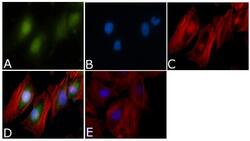
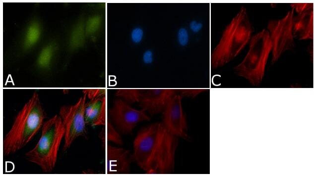
- Immunofluorescent analysis of Aurora A Antibody (35C1)was done on 70% confluent log phase HeLa cells. The cells were fixed with 4% paraformaldehyde for 15 minutes, permeabilized with 0.25% Triton™ X-100 for 10 minutes, and blocked with 5% BSA for 1 hour at room temperature. The cells were labeled with Aurora A Antibody (35C1) (Product # 45-8900) at 1µg/mL in 1% BSA and incubated for 3 hours at room temperature and then labeled with Alexa Fluor 488 Rabbit Anti-Mouse IgG Secondary Antibody (Product # A-11059) at a dilution of 1:400 for 45 minutes at room temperature (Panel a: green). Nuclei (Panel b: blue) were stained with SlowFade® Gold Antifade Mountant with DAPI (Product # S36938). F-actin (Panel c: red) was stained with Alexa Fluor 594 Phalloidin (Product # A12381). Panel d is a merged image showing cytoplasmic and Nuclear localization. Panel e is a no primary antibody control. The images were captured at 40X magnification.
Supportive validation
- Submitted by
- Invitrogen Antibodies (provider)
- Main image
- Experimental details
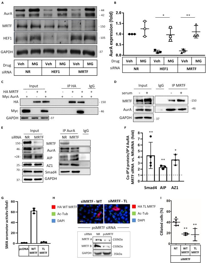
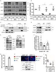
- Figure 3 MRTF interacts with AurA and inhibits its degradation; non-transcriptional effects of MRTF (A) LLC-PK1 cells were transfected with the indicated siRNAs, and 24 hr later placed in serum-free medium supplemented with vehicle (DMSO) or the proteasome inhibitor MG-132 (20 muM) as indicated. After an additional 24 hr, cell lysates were prepared for Western blotting and probed for the indicated proteins. (B) Experiments as in A were quantified by densitometry, and results were normalized to the level of NR-transfected DMSO-treated control is each experiment (n = 3). *p < 0.05, **p < 0.01, the corresponding groups were compared by t test. (C) LLC-PK1 cells were transiently transfected with plasmids encoding HA-tagged MRTF or Myc-tagged AurA or the combinations of these and 24 hr later HA-MRTF was immunoprecipitated with anti-HA. Protein levels in the input and the precipitates were detected by immunoblot using anti-HA or anti-Myc antibodies. (D) Endogenous MRTF was immunoprecipitated from LLC-PK1 cells. Total cell lysates and immunoprecipitates were probed with the indicated antibodies. (E) LLC-PK1 cells were transfected with siNR or siMRTF, and 24 hr later, the level of indicated proteins was examined in the total cell lysates (left) and in immunoprecipitates after pull-down with anti-AurA (right). (F) Quantification of the various protein-protein interactions. The amount of coprecipitating proteins were normalized to the corresponding amount of immunoprecipitated AurA (n