Explore
 Validate
 Learn
Learn Immunohistochemistry
Antibody data
- Antibody Data
- Antigen structure
- References [1]
- Comments [0]
- Validations
- Immunohistochemistry [1]
- Other assay [1]
Submit
Validation data
Reference
Comment
Report error
- Product number
- PA5-37755 - Provider product page

- Provider
- Invitrogen Antibodies
- Product name
- Phospho-TGFBR2 (Ser225, Ser250) Polyclonal Antibody
- Antibody type
- Polyclonal
- Antigen
- Synthetic peptide
- Description
- A suggested positive control for IHC is human brain tissue.
- Reactivity
- Human
- Host
- Rabbit
- Isotype
- IgG
- Vial size
- 100 µL
- Concentration
- 1 mg/mL
- Storage
- -20°C
Submitted references Activation of BK Channels Prevents Hepatic Stellate Cell Activation and Liver Fibrosis Through the Suppression of TGFβ1/SMAD3 and JAK/STAT3 Profibrotic Signaling Pathways.
Yang L, Han B, Zhang M, Wang YH, Tao K, Zhu MX, He K, Zhang ZG, Hou S
Frontiers in pharmacology 2020;11:165
Frontiers in pharmacology 2020;11:165
No comments: Submit comment
Supportive validation
- Submitted by
- Invitrogen Antibodies (provider)
- Main image
- Experimental details
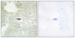
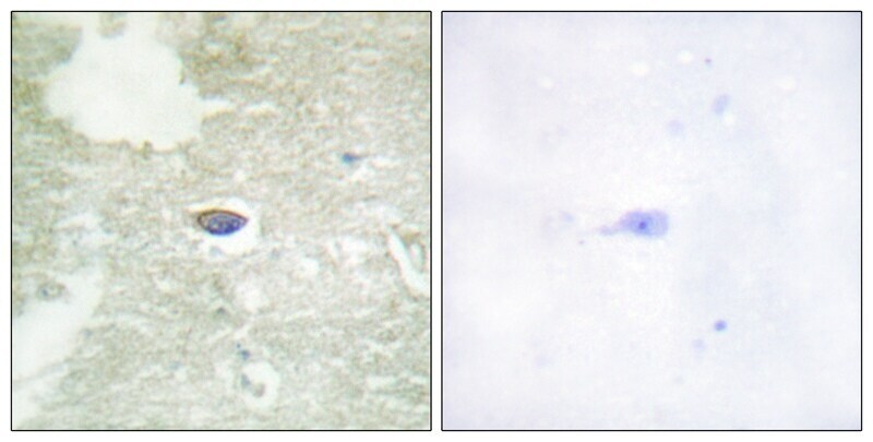
- Immunohistochemical analysis of Phospho-TGFBR2 (Ser225, Ser250) in paraffin-embedded Human brain tissue using Phospho-TGFBR2 (Ser225, Ser250) Polyclonal Antibody (Product # PA5-37755) (left) or the same antibody preincubated with blocking peptide (right).
Supportive validation
- Submitted by
- Invitrogen Antibodies (provider)
- Main image

- Experimental details
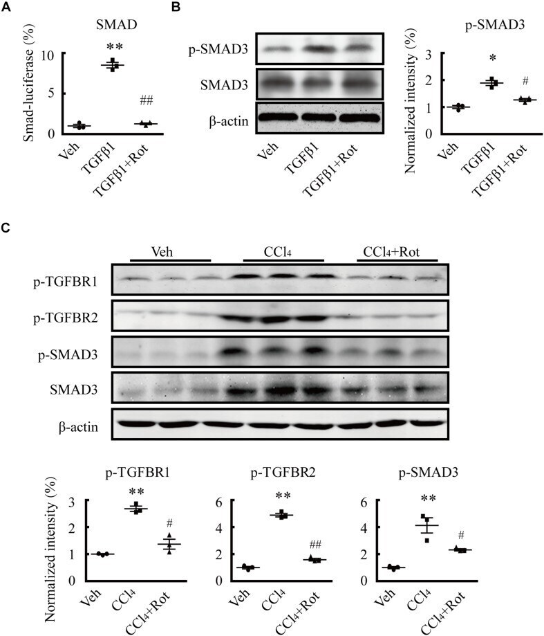
- FIGURE 5 Rottlerin exerts antifibrotic effects through inhibition of the TGFbeta1/SMAD3 signaling pathway. (A) SMAD activity as measured by luciferase reporter assay using reporter vectors PGL4.48[luc2P/SBE/Hygro] in LX2 cells with the indicated treatments. SBE means SMAD binding element (** p < 0.01 compared with the vehicle (Veh) group, ## p < 0.01 compared with the TGFbeta1-treated group; n = 3). (B) Representative western blots ( left ) and normalized intensity ( right ) of the p-SMAD3 levels in primary hepatic stellate cells (HSCs) treated as indicated. Phosphorylation levels of the SMAD3 protein were measured in serum-starved HSCs treated with TGFbeta1 (4 ng/mL) for 1 h. Rottlerin (3 muM) was applied to the medium for 6 h before TGFbeta1 application ( * p < 0.05 compared with the Veh group, # p < 0.05 compared with the TGFbeta1-treated group, n = 3). (C) Representative western blots ( left ) and normalized intensity ( right ) of the levels of total and/or phosphorylated proteins, as indicated, in liver tissues from vehicle, CCl 4 , and rottlerin-treated rats with CCl 4 -induced fibrosis (** p < 0.01 compared with the Veh group, # p < 0.05, and ## p < 0.01 compared with the CCl 4 -treated group, n = 3).