Explore
 Validate
 Learn
Learn711565
antibody from Invitrogen Antibodies
Targeting: TWIST1
ACS3, bHLHa38, BPES2, BPES3, CRS, CRS1, H-twist, SCS, TWIST
 Western blot
Antibody data
- Antibody Data
- Antigen structure
- References [1]
- Comments [0]
- Validations
- Western blot [1]
- Immunocytochemistry [1]
Submit
Validation data
Reference
Comment
Report error
- Product number
- 711565 - Provider product page

- Provider
- Invitrogen Antibodies
- Product name
- TWIST1 Recombinant Polyclonal Antibody
- Antibody type
- Polyclonal
- Antigen
- Other
- Description
- This antibody is predicted to react with Monkey, Bovine, Horse, Mouse
- Concentration
- 0.5 mg/mL
Submitted references Homophilic ATP1A1 binding induces activin A secretion to promote EMT of tumor cells and myofibroblast activation.
Chen YI, Chang CC, Hsu MF, Jeng YM, Tien YW, Chang MC, Chang YT, Hu CM, Lee WH
Nature communications 2022 May 26;13(1):2945
Nature communications 2022 May 26;13(1):2945
No comments: Submit comment
Supportive validation
- Submitted by
- Invitrogen Antibodies (provider)
- Main image
- Experimental details
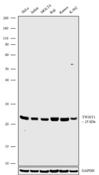
- Western blot analysis was performed on whole cell extracts (30 µg lysate) of HeLa (Lane 1), Jurkat (Lane 2), MOLT4 (Lane 3), Raji (Lane 4), Ramos (Lane 5) and K-562 (Lane 6). The blots were probed with Anti-TWIST1 Recombinant Rabbit Polyclonal Antibody (Product # 711565, 1 µg/mL). A 25 kDa band corresponding to TWIST1 was observed across the cell lines tested. The blots were detected by chemiluminescence using Goat anti-Rabbit IgG (H+L) Superclonal™ Secondary Antibody, HRP conjugate (Product # A27036, 0.4 µg/mL, 1:5000 dilution). Known quantity of protein samples were electrophoresed using Novex® NuPAGE® 4-12% Bis-Tris gel (Product # NP0321BOX), XCell SureLock™ Electrophoresis System (Product # EI0002) and Novex® Sharp Pre-Stained Protein Standard (Product # LC5800). Resolved proteins were then transferred onto a nitrocellulose membrane with iBlot® Dry Blotting System (Product # IB21001). The membrane was probed with the relevant primary and secondary Antibody following blocking with 5% skimmed milk. Chemiluminescent detection was performed using Pierce™ ECL Western blotting Substrate (Product # 32106).

Supportive validation
- Submitted by
- Invitrogen Antibodies (provider)
- Main image
- Experimental details
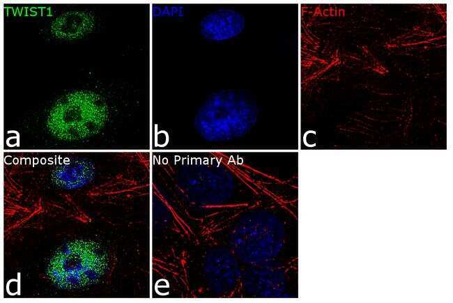
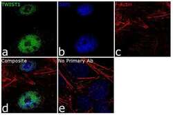
- For immunofluorescence analysis, Ntera2 cells were fixed and permeabilized for detection of endogenous TWIST1 using Anti- TWIST1 Recombinant Rabbit Polyclonal Antibody (Product # 711565, 2 µg/mL) and labeled with Goat anti-Rabbit IgG (H+L) Superclonal™ Secondary Antibody, Alexa Fluor® 488 conjugate (Product # A27034, 1:2000). Panel a) shows representative cells that were stained for detection and localization of TWIST1 protein (green), Panel b) is stained for nuclei (blue) using SlowFade® Gold Antifade Mountant with DAPI (Product # S36938). Panel c) represents cytoskeletal F-actin staining using Rhodamine Phalloidin (Product # R415, 1:300). Panel d) is a composite image of Panels a, b and c clearly demonstrating nuclear localization of TWIST1. Panel e) represents control cells with no primary antibody to assess background. The images were captured at 60X magnification.