 Explore
 Validate
 Learn
Learn Western blot
 ELISA
ELISAAntibody data
- Antibody Data
- Antigen structure
- References [0]
- Comments [0]
- Validations
- Western blot [1]
- Immunohistochemistry [1]
Submit
Validation data
Reference
Comment
Report error
- Product number
- NBP1-77894 - Provider product page

- Provider
- Novus Biologicals
- Proper citation
- Novus Cat#NBP1-77894, RRID:AB_11037437
- Product name
- Rabbit Polyclonal IL-6 Antibody
- Antibody type
- Polyclonal
- Description
- Ion exchange chromatography.
- Reactivity
- Mouse
- Host
- Rabbit
- Isotype
- IgG
- Vial size
- 0.1 mg
- Concentration
- LYOPH
- Storage
- Store lyophilized antibody at 4C. Aliquot reconstituted liquid and store at -20C. Avoid freeze-thaw cycles.
No comments: Submit comment
Supportive validation
- Submitted by
- Novus Biologicals (provider)
- Main image
- Experimental details
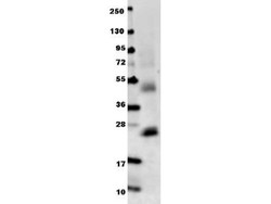
- Western Blot: IL-6 Antibody [NBP1-77894] - Shows detection of recombinant mouse IL-6 raised in E.coli. Recombinant truncated protein (0.1 ug, 21.7 kDa) was loaded on to an SDS-PAGE gel, and after separation, transferred to nitrocellulose. The membrane was blocked with 1% BSA in TBST for 30 min at RT, followed by incubation with Anti-Mouse IL-6 antibody diluted 1:1,000 in 1% BSA in TBST overnight at 4C. After washes, the blot was reacted with secondary antibody DyLight (TM) 649 conjugated Anti-Rabbit IgG (H&L) (Goat) Antibody diluted 1:20,000 in blocking buffer for 30 min. at RT.

Supportive validation
- Submitted by
- Novus Biologicals (provider)
- Main image
- Experimental details
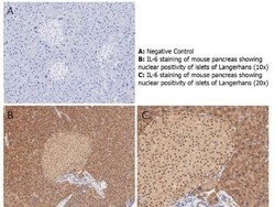
- Immunohistochemistry: IL-6 Antibody [NBP1-77894] - Shows nuclear positivity of islets of Langerhans (brown staining) and cytoplasmic staining in mouse pancreas at 10x and 20x (B & C). Staining was performed on Leica Bond system using the standard protocol. Formalin fixed/paraffin embedded tissue sections were subjected to antigen retrieval with E1 (Leica Microsystems) retrieval solution for 20 min and then incubated with rabbit anti-mouse IL-6 antibody at 1:50 dilution for 60 minutes. Biotinylated Anti-rabbit secondary antibody was used at 1:200 dilution to detect primary antibody. The reaction was developed using streptavidin-HRP conjugated compact polymer system and visualized with chromogen substrate, 3’3-diamino-benzidine substrate (DAB). The sections were then counterstained with hematoxylin to detect cell nuclei.
