Explore
 Validate
 Learn
Learn Western blot
 ELISA
ELISAAntibody data
- Antibody Data
- Antigen structure
- References [2]
- Comments [0]
- Validations
- Western blot [2]
Submit
Validation data
Reference
Comment
Report error
- Product number
- GTX26672 - Provider product page

- Provider
- GeneTex
- Proper citation
- GeneTex Cat#GTX26672, RRID:AB_368308
- Product name
- IL6 antibody
- Antibody type
- Polyclonal
- Reactivity
- Human
- Host
- Rabbit
Submitted references Importance of IL-6, MMP-1, IGF-1, and BAX Levels in Lumbar Herniated Disks and Posterior Longitudinal Ligament in Patients with Sciatic Pain.
MAPK signaling in the quadriceps of patients with chronic obstructive pulmonary disease.
Dagistan Y, Cukur S, Dagistan E, Gezici AR
World neurosurgery 2015 Dec;84(6):1739-46
World neurosurgery 2015 Dec;84(6):1739-46
MAPK signaling in the quadriceps of patients with chronic obstructive pulmonary disease.
Lemire BB, Debigaré R, Dubé A, Thériault ME, Côté CH, Maltais F
Journal of applied physiology (Bethesda, Md. : 1985) 2012 Jul;113(1):159-66
Journal of applied physiology (Bethesda, Md. : 1985) 2012 Jul;113(1):159-66
No comments: Submit comment
Supportive validation
- Submitted by
- GeneTex (provider)
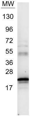
- Main image

- Experimental details
- Western blot using GeneTex anti-IL6 antibody (GTX26672).~1 µg of our control protein recombinant human IL-6 was resolved on a 4-20% Tris-Glycine gel by SDS-PAGE and transferred onto nitrocellulose. The blot shows detection of a band ~21 kDa in size corresponding to anti-IL6 antibody. Molecular weight markers are also shown (MW). After transfer, the membrane was blocked for 30 minutes with 1% BSA-TBST. Detection occurred using peroxidase conjugated anti-Rabbit IgG (GTX27090) secondary antibody diluted 1:40,000 in blocking buffer for 30 min at RT followed by reaction with chemiluminescent substrate.
- Validation comment
- WB
- Submitted by
- GeneTex (provider)
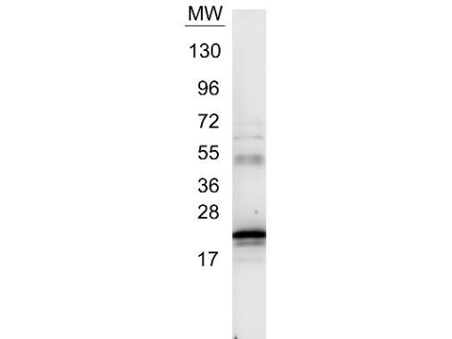
- Main image

- Experimental details
- Western blot using GeneTex anti-IL6 antibody (GTX26672).~1 ?g of our control protein recombinant human IL-6 was resolved on a 4-20% Tris-Glycine gel by SDS-PAGE and transferred onto nitrocellulose. The blot shows detection of a band ~21 kDa in size corresponding to anti-IL6 antibody. Molecular weight markers are also shown (MW). After transfer, the membrane was blocked for 30 minutes with 1% BSA-TBST. Detection occurred using peroxidase conjugated anti-Rabbit IgG (GTX27090) secondary antibody diluted 1:40,000 in blocking buffer for 30 min at RT followed by reaction with chemiluminescent substrate.