 Explore
 Validate
 Learn
Learn Western blot
 ELISA
ELISAAntibody data
- Antibody Data
- Antigen structure
- References [0]
- Comments [0]
- Validations
- Western blot [2]
Submit
Validation data
Reference
Comment
Report error
- Product number
- LF-MA0215 - Provider product page

- Provider
- Invitrogen Antibodies
- Product name
- Anti-ERK1 Monoclonal Antibody (33A5)
- Antibody type
- Monoclonal
- Antigen
- Recombinant full-length protein
- Description
- A suggested positive control for this product is 293T cells.
- Reactivity
- Human, Rat
- Host
- Mouse
- Isotype
- IgG
- Antibody clone number
- 33A5
- Vial size
- 100 µL
- Storage
- -20° C, Avoid Freeze/Thaw Cycles
No comments: Submit comment
Supportive validation
- Submitted by
- Invitrogen Antibodies (provider)
- Main image
- Experimental details
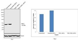
- Knockdown of ERK1 was achieved by transfecting MCF7 cells with ERK1 specific validated siRNAs (Silencer® select Product # s11140).Western blot analysis (Fig a) was performed using whole cell lysates from the ERK1 knock down cells (lane 4), ERK1/ERK2 knock down cells (lane 3), non-specific scrambled siRNA transfected cells (lane 2) and untransfected cells (lane 1). The blots were probed with Anti- ERK1 Mouse Monoclonal Antibody (Product # LF-MA0215, 1:500) and Goat anti-Mouse IgG (H+L) Superclonal™ Secondary Antibody, HRP conjugate (Product # A28177, 0.25 µg/mL, 1:4000 dilution). Densitometric analysis of this western blot is shown in histogram(Fig b). Loss of signal upon siRNA mediated knock down confirms that antibody is specific to ERK1.

- Submitted by
- Invitrogen Antibodies (provider)
- Main image
- Experimental details
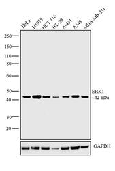
- Western blot analysis was performed on whole cell extracts (30 µg lysate) of HeLa (Lane 1), H-1975 (Lane 2), HCT 116 (Lane 3), HT-29 (Lane 4), A-431 (Lane 5), A549 (Lane 6) and MDA-MB-231 (Lane 7). The blot was probed with Anti-ERK Mouse Monoclonal Antibody (Product # LF-MA0215, 1:2000 dilution) and detected by chemiluminescence using Goat anti-Mouse IgG (H+L) Superclonal™ Secondary Antibody, HRP conjugate (Product # A28177, 0.25 µg/mL, 1:4000 dilution). A 42 kDa band corresponding to ERK was detected across all cell lines tested. Known quantity of protein samples were electrophoresed using Novex® NuPAGE® 4-12 % Bis-Tris gel (Product # NP0321BOX), XCell SureLock™ Electrophoresis System (Product # EI0002) and Novex® Sharp Pre-Stained Protein Standard (Product # LC5800). Resolved proteins were then transferred onto a nitrocellulose membrane with iBlot® 2 Dry Blotting System (Product # IB21001). The membrane was probed with the relevant primary and secondary Antibody following blocking with 5 % skimmed milk. Chemiluminescent detection was performed using Pierce™ ECL Western Blotting Substrate (Product # 32106).
