Explore
 Validate
 Learn
Learn Western blot
 Immunohistochemistry
Antibody data
- Antibody Data
- Antigen structure
- References [3]
- Comments [0]
- Validations
- Immunohistochemistry [11]
- Flow cytometry [1]
Submit
Validation data
Reference
Comment
Report error
- Product number
- NBP1-97712 - Provider product page

- Provider
- Novus Biologicals
- Product name
- Mouse Monoclonal Cytokeratin 19 Antibody
- Antibody type
- Monoclonal
- Description
- Protein A or G purified. The monoclonal antibody specifically reacts with the cytokeratin 19, a 40 kDa polypeptide, which is found in most simple and non-keratinized stratified epithelia. This antibody reacts on formalin fixed, paraffin embedded material and can be used in diagnostic immunopathology. The reaction pattern of cytokeratin 19 is documented in the literature. Antigen origin: human bladder cell line T24. Antigen location: Cytoplasm.
- Reactivity
- Human, Rat, Zebrafish
- Host
- Mouse
- Isotype
- IgG
- Vial size
- 0.1 mg
- Concentration
- 1 mg/ml
- Storage
- Store at 4C short term. Aliquot and store at -20C long term. Avoid freeze-thaw cycles.
Submitted references Expression of Cytokeratin 19 in the epithelial cell of Azo-exposed buccal mucosa.
Impact of the controlled release of a connexin 43 peptide on corneal wound closure in an STZ model of type I diabetes.
A synthetic connexin 43 mimetic peptide augments corneal wound healing.
Handajani J, Hanindriyo L
Medical journal of the Islamic Republic of Iran 2018;32:23
Medical journal of the Islamic Republic of Iran 2018;32:23
Impact of the controlled release of a connexin 43 peptide on corneal wound closure in an STZ model of type I diabetes.
Moore K, Ghatnekar G, Gourdie RG, Potts JD
PloS one 2014;9(1):e86570
PloS one 2014;9(1):e86570
A synthetic connexin 43 mimetic peptide augments corneal wound healing.
Moore K, Bryant ZJ, Ghatnekar G, Singh UP, Gourdie RG, Potts JD
Experimental eye research 2013 Oct;115:178-88
Experimental eye research 2013 Oct;115:178-88
No comments: Submit comment
Supportive validation
- Submitted by
- Novus Biologicals (provider)
- Main image

- Experimental details
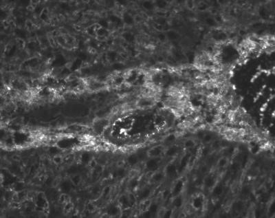
- Immunohistochemistry-Frozen: Cytokeratin 19 Antibody (RCK108) [NBP1-97712] - Analysis using the Unpurified version of NBP1-97712. Immunohistochemistry on frozen section of human liver bile duct epithelium

- Submitted by
- Novus Biologicals (provider)
- Main image
- Experimental details
- Immunohistochemistry: Cytokeratin 19 Antibody (RCK108) [NBP1-97712] - Analysis using the Unpurified version of NBP1-97712. Staining of Human Tongue.
- Submitted by
- Novus Biologicals (provider)
- Main image
- Experimental details
- Immunohistochemistry: Cytokeratin 19 Antibody (RCK108) [NBP1-97712] - Analysis using the Unpurified version of NBP1-97712. Staining of Rat Liver.
- Submitted by
- Novus Biologicals (provider)
- Main image
- Experimental details
- Immunohistochemistry: Cytokeratin 19 Antibody (RCK108) [NBP1-97712] - Analysis using the Unpurified version of NBP1-97712. Staining of Rat Liver.

- Submitted by
- Novus Biologicals (provider)
- Main image

- Experimental details
- Immunohistochemistry: Cytokeratin 19 Antibody (RCK108) [NBP1-97712] - Analysis using the Unpurified version of NBP1-97712. Staining of Rat.
- Submitted by
- Novus Biologicals (provider)
- Main image

- Experimental details
- Immunohistochemistry: Cytokeratin 19 Antibody (RCK108) [NBP1-97712] - Analysis using the Unpurified version of NBP1-97712. Staining of Rat.

- Submitted by
- Novus Biologicals (provider)
- Main image

- Experimental details
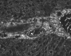
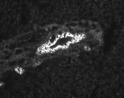
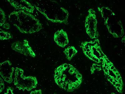
- Immunohistochemistry-Frozen: Cytokeratin 19 Antibody (RCK108) [NBP1-97712] - Immunohistochemistry on a frozen tissue sections of human kidney with NBP1-97712 (RCK108; dilution 1:500).
- Submitted by
- Novus Biologicals (provider)
- Main image

- Experimental details
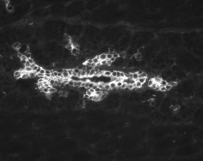
- Immunohistochemistry-Frozen: Cytokeratin 19 Antibody (RCK108) [NBP1-97712] - Immunohistochemistry on frozen tissue sections of human colon with NBP1-97712 (RCK108; dilution 1:500).
- Submitted by
- Novus Biologicals (provider)
- Main image
- Experimental details
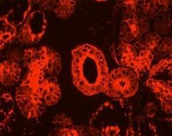
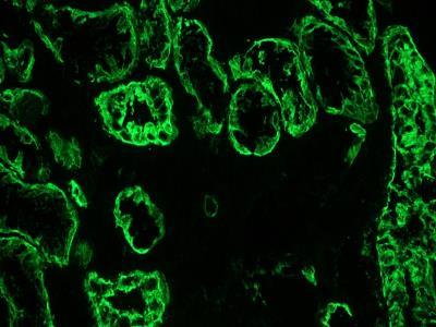
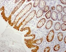
- Immunohistochemistry-Paraffin: Cytokeratin 19 Antibody (RCK108) [NBP1-97712] - Indirect immunoperoxidase staining of human small intestine paraffin tissue section with NBP1-97712 (RCK108; Mouse anti Cytokeratin 19). Dilution 1:50 and microwave pretreatment. Specific staining of the epithelial cells. No reactivity in the connective tissues.

- Submitted by
- Novus Biologicals (provider)
- Main image
- Experimental details
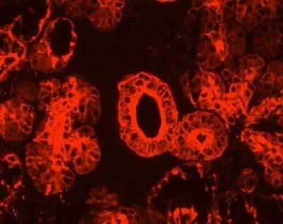
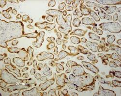
- Immunohistochemistry-Paraffin: Cytokeratin 19 Antibody (RCK108) [NBP1-97712] - Indirect immunoperoxidase staining of human placenta paraffin tissue section with NBP1-97712 (RCK108; Mouse anti Cytokeratin 19). Dilution 1:50 and microwave pretreatment. Specific staining of the epithelial cells. No reactivity in the connective tissues.

- Submitted by
- Novus Biologicals (provider)
- Main image

- Experimental details
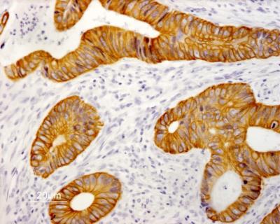
- Immunohistochemistry-Paraffin: Cytokeratin 19 Antibody (RCK108) [NBP1-97712] - Indirect immunoperoxidase staining of human colon adenocarcinoma paraffin tissue section with NBP1-97712 (RCK108; Mouse anti keratin 19). Dilution 1:50 and microwave pretreatment. Specific staining of the epithelial tumor cells. No reactivity in connective tissues.
Supportive validation
- Submitted by
- Novus Biologicals (provider)
- Main image

- Experimental details
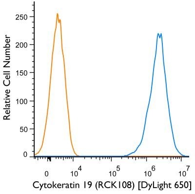
- Flow Cytometry: Cytokeratin 19 Antibody (RCK108) [NBP1-97712] - Flow Cytometry: Cytokeratin 19 Antibody (RCK108) [DyLight 650] [NBP1-97712C] - An intracellular stain was performed on HT-29 cells with Cytokeratin 19 (RCK108) NBP1-97712C (blue) and a matched isotype control NBP1-97005C (orange). Cells were fixed with 4% PFA and then permeablized with 0.1% saponin. Cells were incubated in an antibody dilution of 1 ug/mL for 30 minutes at room temperature. Both antibodies were conjugated to DyLight650. Image using the DyLight 650 form of this antibody.