Explore
 Validate
 Learn
Learn Western blot
Antibody data
- Antibody Data
- Antigen structure
- References [0]
- Comments [0]
- Validations
- Western blot [5]
- Immunocytochemistry [1]
- Immunohistochemistry [2]
Submit
Validation data
Reference
Comment
Report error
- Product number
- MA5-15862 - Provider product page

- Provider
- Invitrogen Antibodies
- Product name
- Cytokeratin 19 Monoclonal Antibody (1H6)
- Antibody type
- Monoclonal
- Antigen
- Purifed from natural sources
- Description
- MA5-15862 targets KRT19 in IHC and WB applications and shows reactivity with Human samples.
- Antibody clone number
- 1H6
- Concentration
- Conc. Not Determined
No comments: Submit comment
Supportive validation
- Submitted by
- Invitrogen Antibodies (provider)
- Main image

- Experimental details
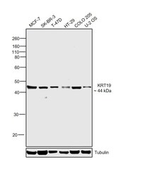
- Western blot analysis of KRT19 using KRT19 monoclonal antibody (Product # MA5-15862) in T47D (1), MCF-7 (2), SKBR-3 (3), HepG2 (4), Caco-2 (5) and SW620 (6) cell lysate.
- Submitted by
- Invitrogen Antibodies (provider)
- Main image

- Experimental details
- Western blot analysis of KRT19 using KRT19 monoclonal antibody (Product # MA5-15862) in T47D (1), MCF-7 (2), SKBR-3 (3), HepG2 (4), Caco-2 (5) and SW620 (6) cell lysate.

- Submitted by
- Invitrogen Antibodies (provider)
- Main image

- Experimental details
- Western blot analysis of KRT19 using KRT19 monoclonal antibody (Product # MA5-15862) in T47D (1), MCF-7 (2), SKBR-3 (3), HepG2 (4), Caco-2 (5) and SW620 (6) cell lysate.

- Submitted by
- Invitrogen Antibodies (provider)
- Main image

- Experimental details
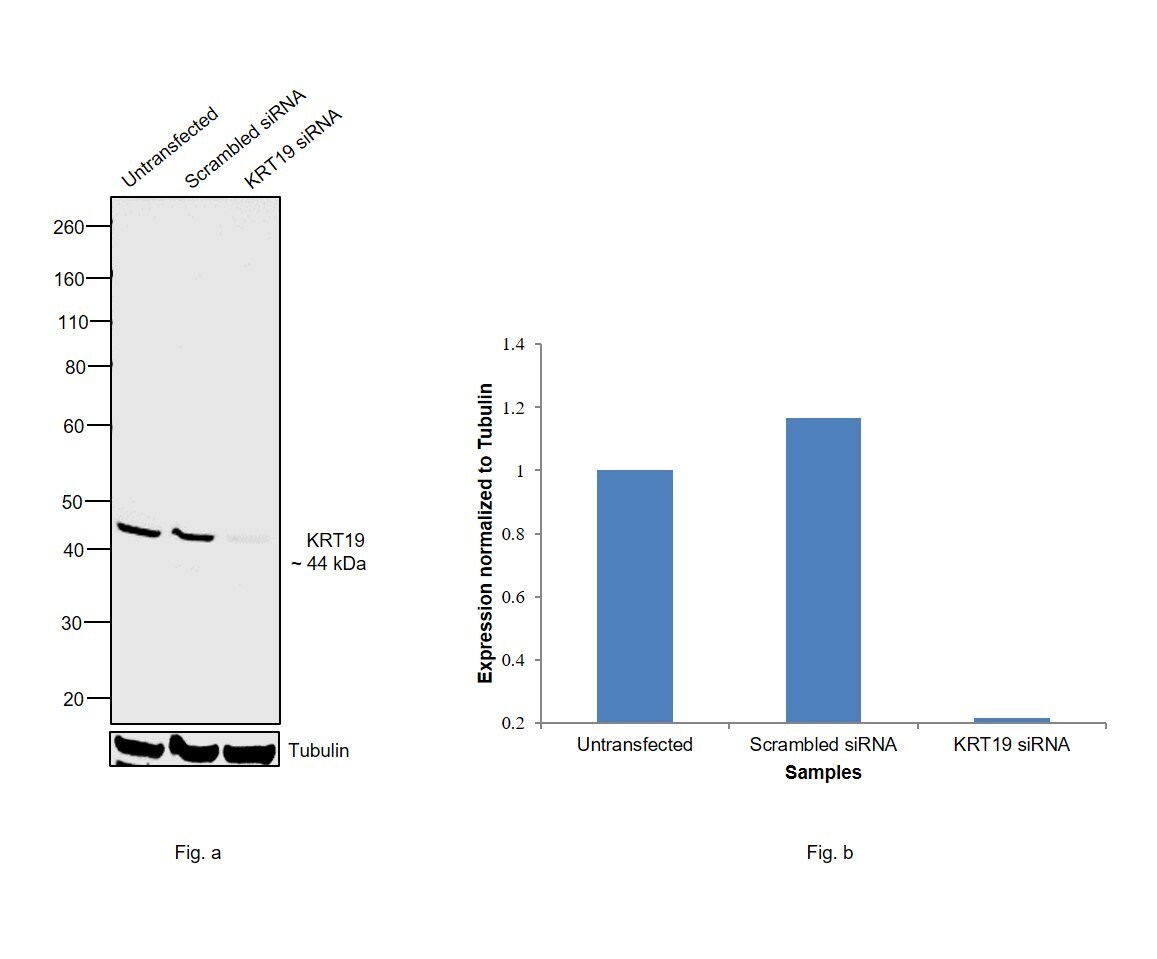
- Knockdown of KRT19 was achieved by transfecting MCF-7 with KRT19 specific siRNAs (Silencer® select Product # s7998). Western blot analysis (Fig. a) was performed using membrane enriched extracts from the KRT19 knockdown cells (lane 3), non-specific scrambled siRNA transfected cells (lane 2) and untransfected cells (lane 1). The blot was probed with Cytokeratin 19 Monoclonal Antibody (1H6) (Product # MA5-15862, 1:1000 dilution) and Goat anti-Mouse IgG (H+L), Superclonal™ Recombinant Secondary Antibody, HRP (Product # A28177, 0.25µg/ml, 1:4000 dilution). Densitometric analysis of this western blot is shown in histogram (Fig. b). Decrease in signal upon siRNA mediated knock down confirms that antibody is specific to KRT19.
- Submitted by
- Invitrogen Antibodies (provider)
- Main image
- Experimental details
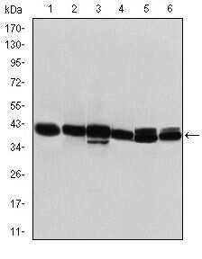
- Western blot was performed using Anti-Cytokeratin 19 Monoclonal Antibody (1H6) (Product # MA5-15862) and a 44 kDa band corresponding to KRT19 was observed across cell lines tested. Membrane enriched extracts (30 µg lysate) of MCF-7 (Lane 1), SK-BR-3 (Lane 2), T-47D (Lane 3), HT-29 (Lane 4), COLO 205 (Lane 5) and U-2 OS (Lane 6) were electrophoresed using NuPAGE™ 4-12% Bis-Tris Protein Gel (Product # NP0322BOX). Resolved proteins were then transferred onto a nitrocellulose membrane (Product # IB23001) by iBlot® 2 Dry Blotting System (Product # IB21001). The blot was probed with the primary antibody (1:1000 dilution) and detected by chemiluminescence with Goat anti-Mouse IgG (H+L) Superclonal™ Recombinant Secondary Antibody, HRP (Product # A28177, 1:4000 dilution) using the iBright FL 1000 (Product # A32752). Chemiluminescent detection was performed using Novex® ECL Chemiluminescent Substrate Reagent Kit (Product # WP20005).

Supportive validation
- Submitted by
- Invitrogen Antibodies (provider)
- Main image
- Experimental details
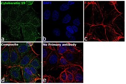
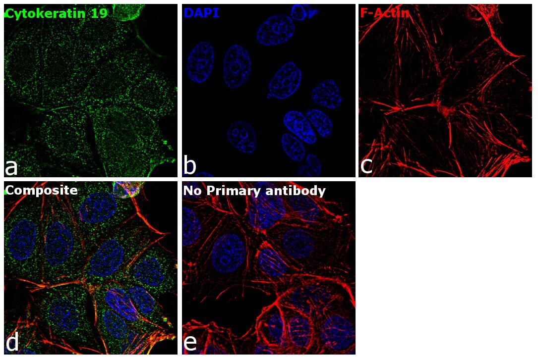
- Immunofluorescence analysis of Cytokeratin 19 was performed using 70% confluent log phase MCF-7 cells. The cells were fixed with 4% paraformaldehyde for 10 minutes, permeabilized with 0.1% Triton™ X-100 for 15 minutes, and blocked with 2% BSA for 1 hour at room temperature. The cells were labeled with Cytokeratin 19 Mouse Monoclonal Antibody (1H6) (Product # MA5-15862) at 1:100 dilution in 0.1% BSA, incubated at 4 degree Celsius overnight and then with Goat anti-Mouse IgG (H+L) Superclonal™ Recombinant Secondary Antibody, Alexa Fluor® 488 conjugate (Product # A28175) at a dilution of 1:2000 for 45 minutes at room temperature (Panel a: green). Nuclei (Panel b: blue) were stained with ProLong™ Diamond Antifade Mountant with DAPI (Product # P36962). F-actin (Panel c: red) was stained with Rhodamine Phalloidin (Product # R415, 1:300). Panel d represents the merged image showing cytoplasmic localization. Panel e represents control cells with no primary antibody to assess background. The images were captured at 60X magnification.
Supportive validation
- Submitted by
- Invitrogen Antibodies (provider)
- Main image

- Experimental details
- Immunohistochemical analysis of paraffin-embedded rectum cancer tissues (left) and lung cancer tissues (right) using KRT19 monoclonal antibody (Product # MA5-15862) followed with DAB staining.

- Submitted by
- Invitrogen Antibodies (provider)
- Main image

- Experimental details
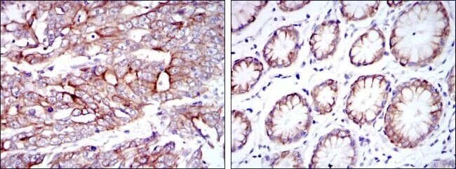
- Immunohistochemical analysis of paraffin-embedded stomach cancer tissues (left) and stomach tissues (right) using KRT19 monoclonal antibody (Product # MA5-15862) followed with DAB staining.