 Explore
 Validate
 Learn
Learn Western blot
Antibody data
- Antibody Data
- Antigen structure
- References [2]
- Comments [0]
- Validations
- Western blot [5]
- Immunocytochemistry [2]
- Immunohistochemistry [4]
- Other assay [2]
Submit
Validation data
Reference
Comment
Report error
- Product number
- MA5-15884 - Provider product page

- Provider
- Invitrogen Antibodies
- Product name
- Cytokeratin 19 Monoclonal Antibody (4E8)
- Antibody type
- Monoclonal
- Antigen
- Purifed from natural sources
- Description
- MA5-15884 targets KRT19 in IF, IHC, and WB applications and shows reactivity with Human samples.
- Antibody clone number
- 4E8
- Concentration
- Conc. Not Determined
Submitted references Tetrahydroxylated bile acids improve cholestatic liver and bile duct injury in the Mdr2(-/-) mouse model of sclerosing cholangitis via immunomodulatory effects.
Ulipristal Acetate Interferes With Actin Remodeling Induced by 17β-Estradiol and Progesterone in Human Endometrial Stromal Cells.
Fuchs CD, Dixon ED, Hendrikx T, Mlitz V, Wahlström A, Ståhlman M, Scharnagl H, Stojakovic T, Binder CJ, Marschall HU, Trauner M
Hepatology communications 2022 Sep;6(9):2368-2378
Hepatology communications 2022 Sep;6(9):2368-2378
Ulipristal Acetate Interferes With Actin Remodeling Induced by 17β-Estradiol and Progesterone in Human Endometrial Stromal Cells.
Shortrede JE, Montt-Guevara MM, Pennacchio G, Finiguerra M, Giannini A, Genazzani AD, Simoncini T
Frontiers in endocrinology 2018;9:350
Frontiers in endocrinology 2018;9:350
No comments: Submit comment
Supportive validation
- Submitted by
- Invitrogen Antibodies (provider)
- Main image

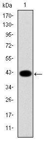
- Experimental details
- Western blot analysis of KRT19 using a KRT19 monoclonal antibody (Product # MA5-15884) against a human KRT19 (AA: 115-269) recombinant protein.

- Submitted by
- Invitrogen Antibodies (provider)
- Main image

- Experimental details
- Western blot analysis of KRT19 using a KRT19 monoclonal antibody (Product # MA5-15884) against a human KRT19 (AA: 115-269) recombinant protein.
- Submitted by
- Invitrogen Antibodies (provider)
- Main image
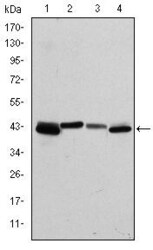
- Experimental details
- Western blot analysis of KRT19 using KRT19 monoclonal antibody (Product # MA5-15884) in T47D (1), MCF-7 (2), HepG2 (3) and SW620 (4) cell lysate.

- Submitted by
- Invitrogen Antibodies (provider)
- Main image

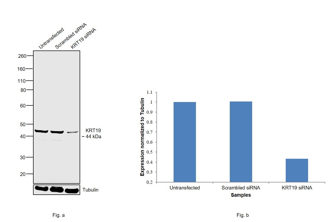
- Experimental details
- Knockdown of KRT19 was achieved by transfecting MCF-7 with KRT19 specific siRNAs (Silencer® select Product # s7998). Western blot analysis (Fig. a) was performed using membrane enriched extracts from the KRT19 knockdown cells (lane 3), non-specific scrambled siRNA transfected cells (lane 2) and untransfected cells (lane 1). The blot was probed with Cytokeratin 19 Monoclonal Antibody (4E8) (Product # MA5-15884, 1:1000 dilution) and Goat anti-Mouse IgG (H+L), Superclonal™ Recombinant Secondary Antibody, HRP (Product # A28177, 0.25µg/ml, 1:4000 dilution). Densitometric analysis of this western blot is shown in histogram (Fig. b). Decrease in signal upon siRNA mediated knock down confirms that antibody is specific to KRT19.
- Submitted by
- Invitrogen Antibodies (provider)
- Main image
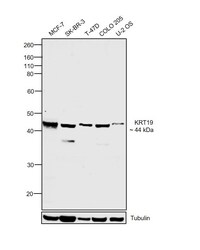
- Experimental details
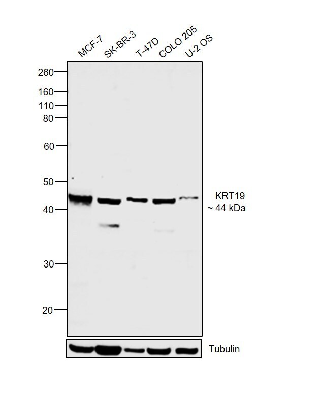
- Western blot was performed using Anti-Cytokeratin 19 Monoclonal Antibody (4E8) (Product # MA5-15884) and a 44 kDa band corresponding to KRT19 was observed across cell lines tested. Membrane enriched extracts (30 µg lysate) of MCF-7 (Lane 1), SK-BR-3 (Lane 2), T-47D (Lane 3), COLO 205 (Lane 4) and U-2 OS (Lane 5) were electrophoresed using NuPAGE™ 4-12% Bis-Tris Protein Gel (Product # NP0322BOX). Resolved proteins were then transferred onto a nitrocellulose membrane (Product # IB23001) by iBlot® 2 Dry Blotting System (Product # IB21001). The blot was probed with the primary antibody (1:1000 dilution) and detected by chemiluminescence with Goat anti-Mouse IgG (H+L) Superclonal™ Recombinant Secondary Antibody, HRP (Product # A28177, 1:4000 dilution) using the iBright FL 1000 (Product # A32752). Chemiluminescent detection was performed using Novex® ECL Chemiluminescent Substrate Reagent Kit (Product # WP20005).
Supportive validation
- Submitted by
- Invitrogen Antibodies (provider)
- Main image

- Experimental details
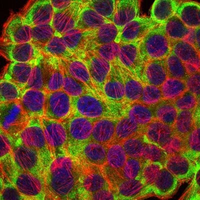
- Immunofluorescence analysis of HepG2 cells using KRT19 monoclonal antibody (Product # MA5-15884) (Green). Blue: DRAQ5 fluorescent DNA dye. Red: actin filaments have been labeled with phalloidin.
- Submitted by
- Invitrogen Antibodies (provider)
- Main image
- Experimental details
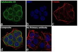
- Immunofluorescence analysis of Cytokeratin 19 was performed using 70% confluent log phase MCF-7 cells. The cells were fixed with 4% paraformaldehyde for 10 minutes, permeabilized with 0.1% Triton™ X-100 for 15 minutes, and blocked with 2% BSA for 1 hour at room temperature. The cells were labeled with Cytokeratin 19 Mouse Monoclonal Antibody (4E8) (Product # MA5-15884) at 1:100 dilution in 0.1% BSA, incubated at 4 degree Celsius overnight and then with Goat anti-Mouse IgG (H+L) Superclonal™ Recombinant Secondary Antibody, Alexa Fluor® 488 conjugate (Product # A28175) at a dilution of 1:2000 for 45 minutes at room temperature (Panel a: green). Nuclei (Panel b: blue) were stained with ProLong™ Diamond Antifade Mountant with DAPI (Product # P36962). F-actin (Panel c: red) was stained with Rhodamine Phalloidin (Product # R415, 1:300). Panel d represents the merged image showing cytoplasmic localization. Panel e represents control cells with no primary antibody to assess background. The images were captured at 60X magnification.

Supportive validation
- Submitted by
- Invitrogen Antibodies (provider)
- Main image
- Experimental details
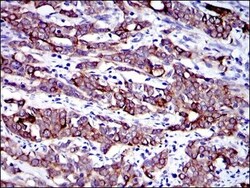
- Immunohistochemical analysis of paraffin-embedded human cervical cancer tissues using KRT19 monoclonal antibody (Product # MA5-15884) followed with DAB staining.

- Submitted by
- Invitrogen Antibodies (provider)
- Main image
- Experimental details
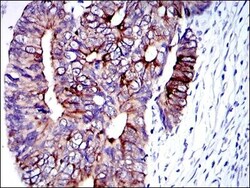
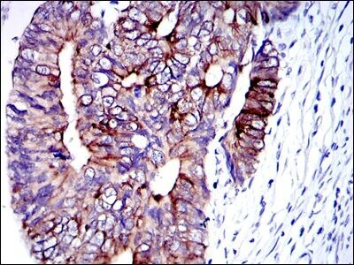
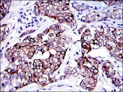
- Immunohistochemical analysis of paraffin-embedded human colon cancer tissues using KRT19 monoclonal antibody (Product # MA5-15884) followed with DAB staining.
- Submitted by
- Invitrogen Antibodies (provider)
- Main image
- Experimental details
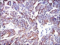
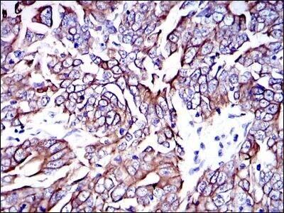
- Immunohistochemical analysis of paraffin-embedded human stomach cancer tissues using KRT19 monoclonal antibody (Product # MA5-15884) followed with DAB staining.
- Submitted by
- Invitrogen Antibodies (provider)
- Main image

- Experimental details
- Immunohistochemical analysis of paraffin-embedded human bladder cancer tissues using KRT19 monoclonal antibody (Product # MA5-15884) followed with DAB staining.
Supportive validation
- Submitted by
- Invitrogen Antibodies (provider)
- Main image
- Experimental details
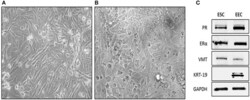
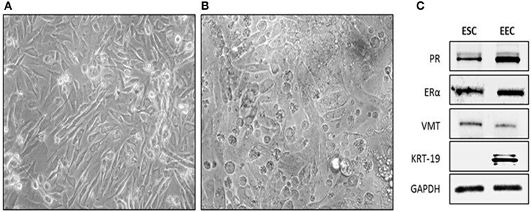
- Figure 1 Isolation and characterization of human endometrial cells. Cells were obtained from endometrial biopsies and cultured in DMEM-F12 supplemented with 10% FBS. (A) Phase contrast microscopy of Endometrial Stromal Cells (ESC), cells appeared flattened and elongated with a fibroblast-like shape. Photo taken 4 day after plating (20X). (B) Phase contrast microscopy of EEC cells appeared cuboidal. Photo was taken 4 days after plating. (C) Representative western-blot blot of Vimentin (VMT), Cytokeratin 19 (KRT-19), Estrogen Receptor alpha (ERalpha), Progesterone Receptor (PR), and GAPDH. ESC were VMT/ERalpha/PR positive. EEC were KRT-19/ERalpha/PR positive. GAPDH was used as loading control.
- Submitted by
- Invitrogen Antibodies (provider)
- Main image
- Experimental details
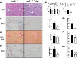
- 4 FIGURE Tetrahydroxylated bile acid (THBA) feeding improves liver and bile duct injury in the Mdr24 -/- mouse model of sclerosing cholangitis. (A) Representative H&E images (x10 magnification) with improved liver histology in Mdr2 -/- mice fed a 0.5% wt/wt THBA-enriched diet for 4 weeks. (B) Serum biochemistry reflects reduced levels of transaminases (ALT, AST) Mdr2 -/- mice fed 0.5% wt/wt THBA. Total bile acid (BA) levels as well as ALP levels tended to be reduced due to THBA feeding. (C) Representative MAC-2 immunohistochemistry images (x10 magnification) showing reduced numbers of macrophages in the livers of THBA-fed Mdr2 -/- mice. (D) Real-time PCR was used to assess the mRNA expression of F4/80 , as markers of inflammation, which was reduced in THBA-fed Mdr2 -/- mice. (E) Representative sirius red stainings (x10 magnification) show tendentially reduced biliary fibrosis in Mdr2 -/- mice fed 0.5% wt/wt THBA. (F) Real-time PCR was used to assess the mRNA expression of fibrotic marker collagen type I alpha 1 ( Col1a1) , which was reduced in THBA-fed Mdr2 -/- mice. (G) Representative CK19 immunohistochemistry pictures (x10 magnification) show reduced cholangiocyte proliferation in THBA-fed Mdr2 -/- mice. (H) Real-time PCR was used to assess the mRNA expression of cholangiocyte proliferation marker CK19 , which was reduced in THBA-fed Mdr2 -/- mice. mRNA expression values were normalized against 36b4 levels and are shown relative to expression level in WT controls. *Signific
